

Start your One-Week Free Trial
Already subscribed? Log in »

Tracheobronchial Tree
Here we’ll learn about the tracheobronchial tree, which comprises conducting and respiratory structures.
 Ultimately, bronchial divisions give rise to conducting bronchioles, in which the cartilaginous plates are completely absent.
The final generation of conducting bronchioles are called terminal bronchioles; they mark the end of the conducting portion of the respiratory tract.
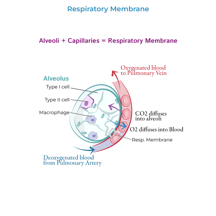
Respiratory bronchioles have alveoli sprouting from them; alveoli are the thin-walled sacs where gas exchange occurs.
Respiratory bronchioles give rise to several alveolar ducts, which are lined with alveoli that open to alveolar sacs.
We’ve cut open an alveolar sac so that you can see that air passes through the ducts and enters the alveoli;
Alveolar pores facilitate pressure equalization between the alveoli.
The alveoli are in close contact with pulmonary capillary beds, which is where gas exchange occurs.
Ultimately, bronchial divisions give rise to conducting bronchioles, in which the cartilaginous plates are completely absent.
The final generation of conducting bronchioles are called terminal bronchioles; they mark the end of the conducting portion of the respiratory tract.
Respiratory bronchioles have alveoli sprouting from them; alveoli are the thin-walled sacs where gas exchange occurs.
Respiratory bronchioles give rise to several alveolar ducts, which are lined with alveoli that open to alveolar sacs.
We’ve cut open an alveolar sac so that you can see that air passes through the ducts and enters the alveoli;
Alveolar pores facilitate pressure equalization between the alveoli.
The alveoli are in close contact with pulmonary capillary beds, which is where gas exchange occurs.
 Bronchial arteries and veins supply systemic blood to the bronchial tree. The arteries arise from the thoracic aorta and its branches.
Venous blood pathways differ on the right and left sides, and display more individual variation. It is commonly reported that blood is returned via the azygos vein on the right and the hemiazygos vein on the left; some blood may also be returned via the pulmonary veins, which creates a shunt.
The airways are innervated by branches of the pulmonary plexuses; vagus nerves constrict the bronchioles, and sympathetic fibers dilate them.
The subpleural plexus drains the lungs of lymphatic fluid; the plexus is subdivided into deep and superficial groups, which communicate peripherally.
The deep group travels within the bronchial submucosa and peribronchial connective tissues; these vessels follow the pulmonary artery and vein and the bronchi.
The superficial lymph vessels drain the lung tissue and visceral pleura; they converge at hilum and drain into bronchopulmonary lymph nodes.
Lung cancer often originates in the bronchi.
Pulmonary embolism (aka PE) obstructs arterial supply. In a PE, gas exchange is reduced, and blood oxygen levels drop.
Chronic Bronchitis & Emphysema
Asthma
Bronchiectasis
Bronchial arteries and veins supply systemic blood to the bronchial tree. The arteries arise from the thoracic aorta and its branches.
Venous blood pathways differ on the right and left sides, and display more individual variation. It is commonly reported that blood is returned via the azygos vein on the right and the hemiazygos vein on the left; some blood may also be returned via the pulmonary veins, which creates a shunt.
The airways are innervated by branches of the pulmonary plexuses; vagus nerves constrict the bronchioles, and sympathetic fibers dilate them.
The subpleural plexus drains the lungs of lymphatic fluid; the plexus is subdivided into deep and superficial groups, which communicate peripherally.
The deep group travels within the bronchial submucosa and peribronchial connective tissues; these vessels follow the pulmonary artery and vein and the bronchi.
The superficial lymph vessels drain the lung tissue and visceral pleura; they converge at hilum and drain into bronchopulmonary lymph nodes.
Lung cancer often originates in the bronchi.
Pulmonary embolism (aka PE) obstructs arterial supply. In a PE, gas exchange is reduced, and blood oxygen levels drop.
Chronic Bronchitis & Emphysema
Asthma
Bronchiectasis
Trachea & Bronchi
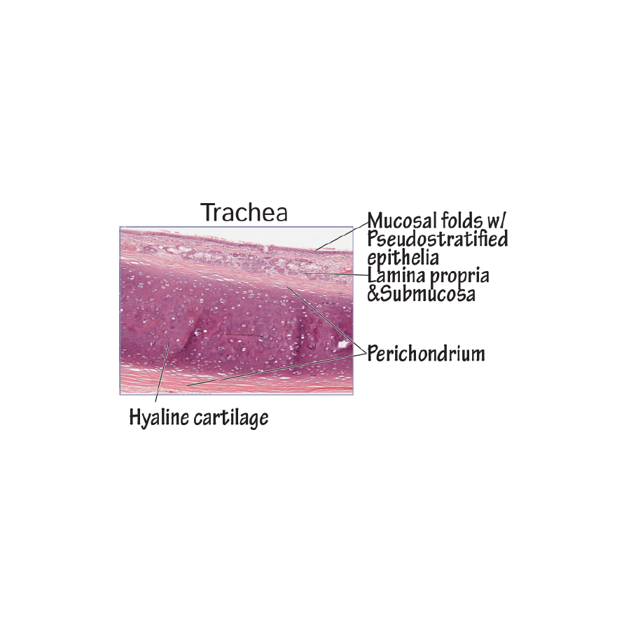
Trachea
The trachea attaches to the cricoid cartilage of the larynx at level C 6 and descends through the neck and into the mediastinum, where it bifurcates into left and right main, aka primary bronchi, at the sternal angle.
The trachea comprises 16-20 c-shaped rings made of hyaline cartilage, which hold it open circumferentially and provides a limited degree of flexibility and mobility. The posterior wall comprises a longitudinal strip of smooth muscle, called the trachealis muscle.
Cross Section
We draw a single c-ring cartilage in cross section and we indicate the mucosal lining with glands, elastic fibers, and blood vessels.
The trachealis muscle closing the open part of the C-ring, and, for context, indicate that the esophagus runs posteriorly to it.
Bronchi
The right and left main bronchi enter the root of the lungs at their hila.
The right main bronchus is generally shorter, wider, and more vertically oriented than the left bronchus, so inspirated foreign objects tend to lodge on the right side.
After entering the lungs, the main bronchi divide into lobar (aka secondary) bronchi, each of which supplies a specific lobe of the lungs; recall that the right lung has three lobes, the left has two.
The lobar bronchi divide into segmental (aka tertiary) bronchi, which supply bronchopulmonary segments of the lungs.
Notice that as we move down the bronchial tree, the cartilaginous plates become increasingly irregular.
Bronchioles
Neurovasculature
*Clinical Correlations