Start your One-Week Free Trial
Already subscribed? Log in »
Respiratory System Anatomy (Overview)
Functions of the Respiratory System
Functional divisions:
The conducting portion is the portion that “conducts” the air and includes the nose, nasal cavity, pharynx, larynx, trachea, bronchi, and bronchioles.
The respiratory portion is the portion that participates in gas exchange and includes the respiratory bronchioles, alveolar ducts and sacs, and the alveoli themselves.
Respiratory anatomy
The nasal cavity
Divided into right and left halves by the nasal septum and further subdivided by nasal conchae.
Paranasal Sinuses
Hollow spaces in the bones; named according to the bones.
The frontal sinus is in the forehead.
The ethmoidal air cells are small cavities collectively called the ethmoid sinus, lie between the eyes and behind the bridge of the nose.
Maxillary sinus is a space in each cheekbone.
Sphenoid sinus in the sphenoid bone.
The paranasal sinuses communicate with the nasal cavity, and their mucosal linings help to warm the inspired air and trap particles.
Unfortunately, infection can spread from the nasal cavity to the paranasal sinuses, leading to a sinus infection.
Palate
The palate, which comprises bony and soft tissues, is below the nasal cavity.
Oral Cavity
The oral cavity, which is bound anteriorly by the lips and teeth; inferiorly, it is bound by the floor of the mouth. The tongue is attached to the floor of the mouth and takes up most of the space of the oral cavity.
Hyoid bone
Is a bone suspended in the neck that supports the tongue and provides attachment sites for the muscles used in swallowing, breathing, and speech.
We commonly call this the “voice box” because we use it to produce sounds.
The thyroid cartilage is the large cartilage that contributes to the anterior and lateral sides of the larynx. Its name describes its shield-like shape (the Greek word for “shield-like is “thyreos”).
The epiglottis attaches to the inside of the thyroid cartilage and projects superiorly. The epiglottis is a leaf-shaped piece of cartilage that closes off the larynx during swallowing to prevent foods and liquids from entering the larynx.
Vestibular and vocal folds (aka vocal cords); the vocal folds are responsible for sound production. Be aware that the vestibular, aka ventricular folds are sometimes referred to as the “false vocal cords.”
A continuous muscular tube-like structure that provides passageway for air, foods, and liquids. The pharynx is what we commonly refer to as “the throat.”
Where the pharynx is open to the nasal cavity, we call it the nasopharynx; thanks to the soft palate, only air passes through here, except when something goes “up your nose.”
Where the pharynx is open to the oral cavity, we call it the oropharynx, and, where it’s open to the larynx, we call it the laryngopharynx. Air, foods, and liquids pass through these two sections of the pharynx.
Below the laryngopharynx, the pharynx transitions to become the esophagus, a structure of the digestive system.
Trachea & Bronchi
Commonly called the windpipe, the trachea begins at the distal end of the larynx and enters the mediastinum.
This tube comprises 16-20 c-shaped rings of cartilage connected by annular ligaments; the trachealis muscle forms the posterior wall of the tube.
At its distal end, the trachea bifurcates to form the right and left main, aka, primary bronchi.
The main bronchi enter the lungs and continue to branch into progressively smaller passageways as follows: lobar, aka secondary bronchi; segmental, aka tertiary bronchi; and then into a series of bronchioles.
Terminal bronchioles give rise to respiratory bronchioles; this marks the transition between conducting and respiratory portions of the respiratory tract.
The smallest respiratory bronchioles give rise to alveolar ducts, at the end of which we find alveolar sacs. Along this final passageway, small, thin-walled sacs of epithelia called alveoli provide the surface for gas exchange with nearby pulmonary capillary beds.
Lungs
The right lung is slightly larger, and has three lobes, which are named for their relative positions: superior, middle, and inferior lobes.
The left lung is slightly smaller and has only two lobes, superior and inferior. Indicate that the indentation in the left lung is called the cardiac notch; this is where the heart sits.
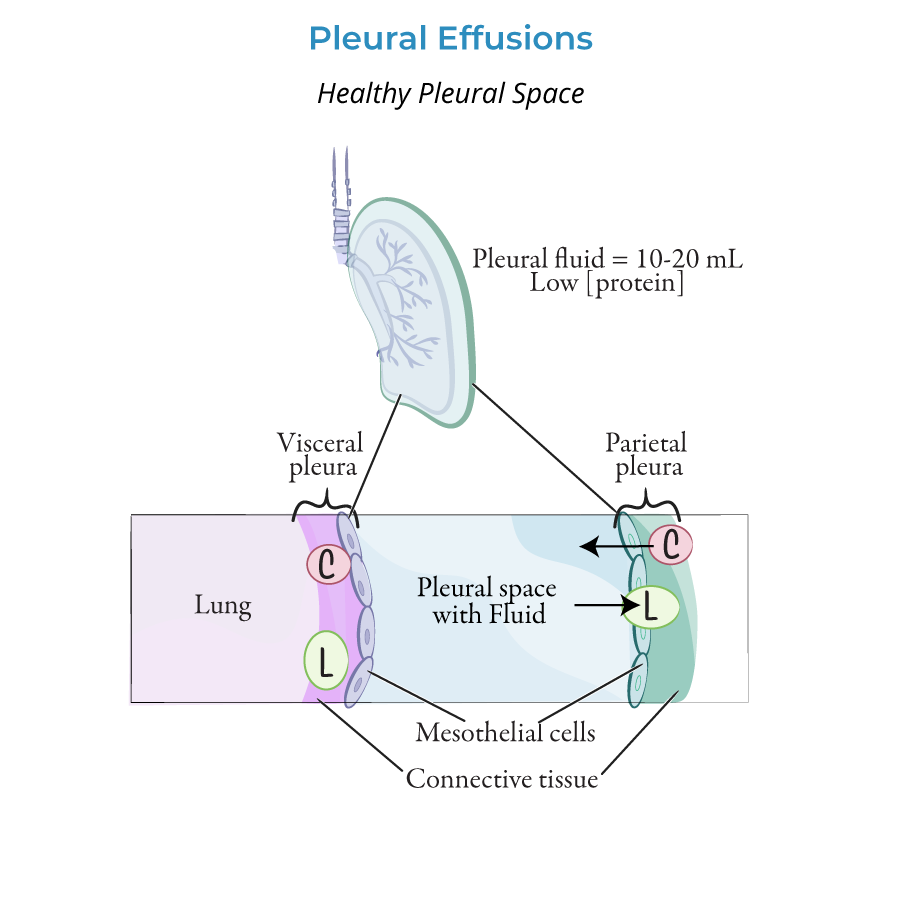
The lungs are covered in a double-layered serous membrane, called pleura, separated by a thin layer of fluid, which helps to reduce friction during breathing.
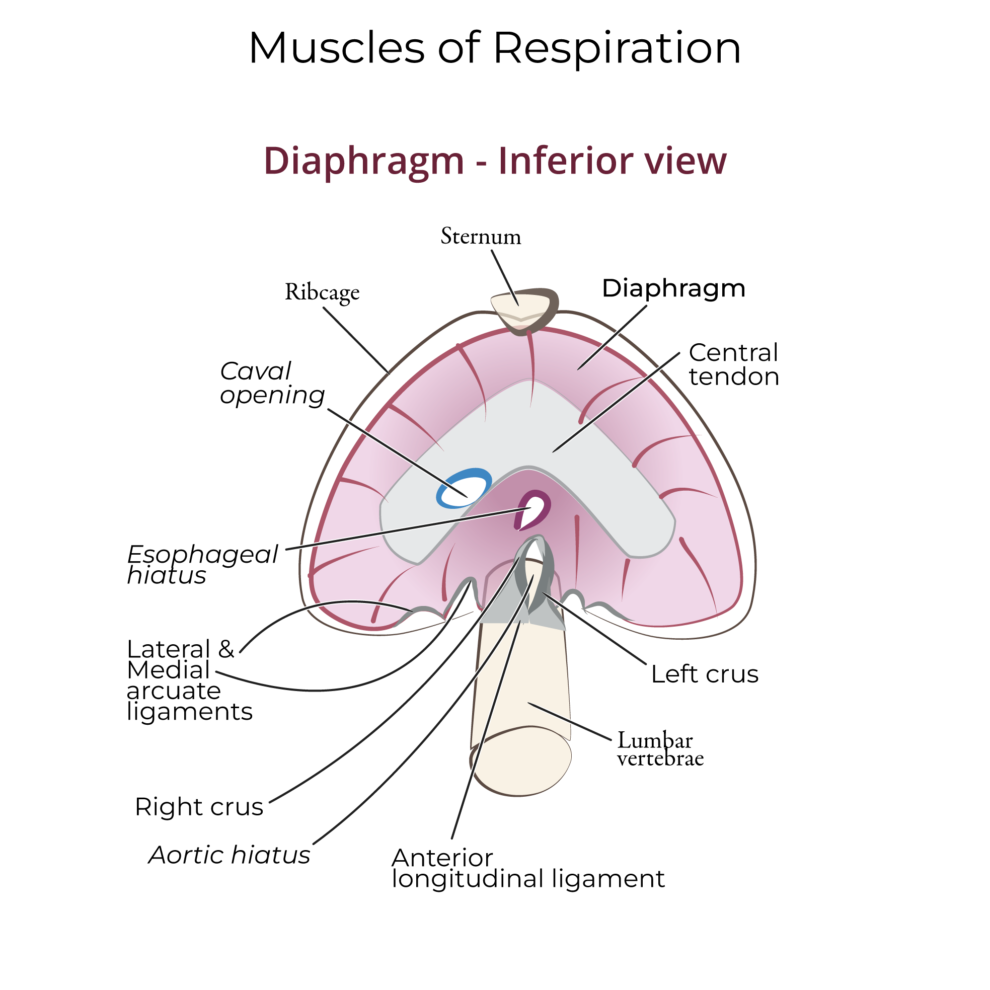
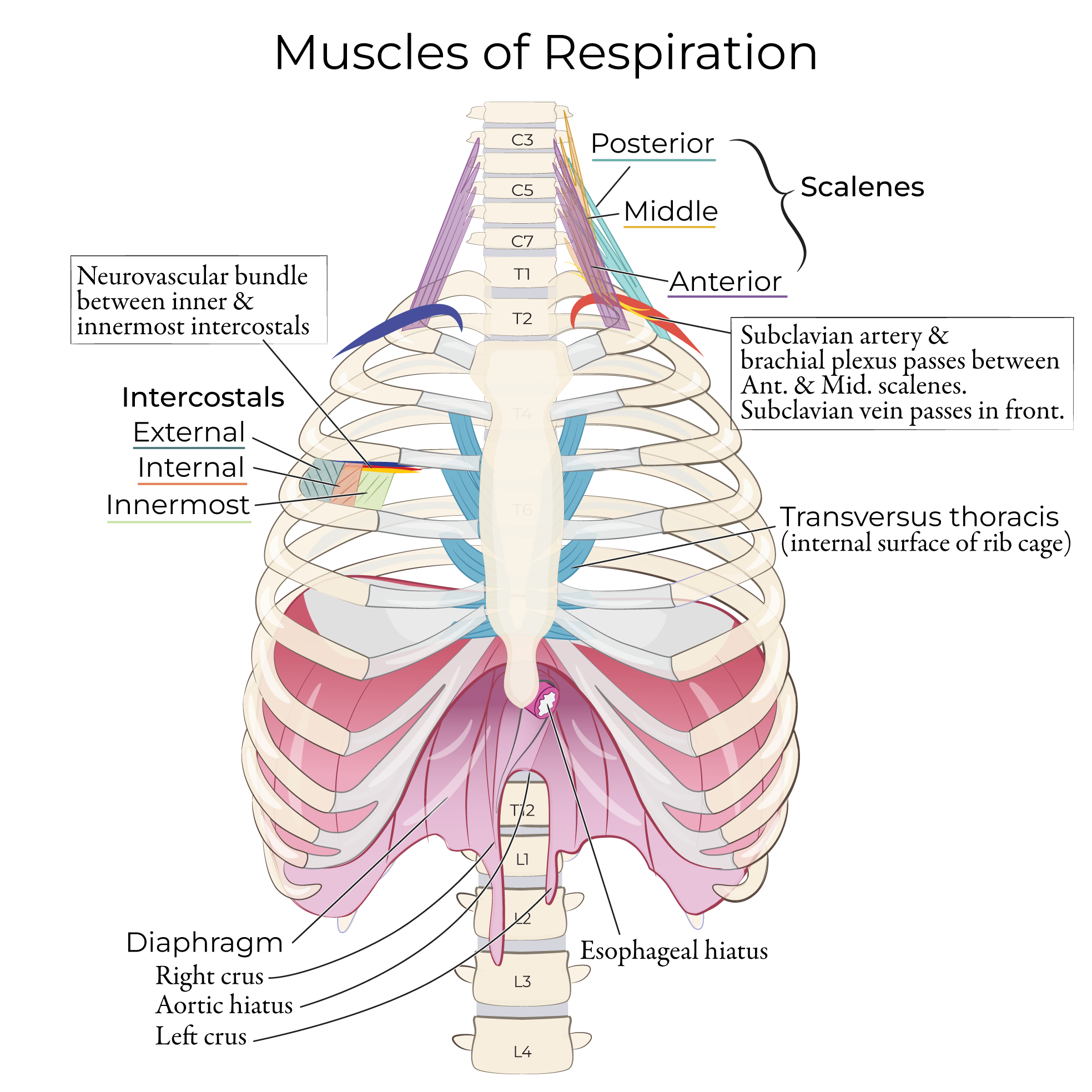
 Below the lungs, indicate the diaphragm. This thin, dome-shaped muscle expands and contracts to facilitate changes in the dimensions of the thoracic cavity, which in turn drives air movement into and out of the lungs.