

Start your One-Week Free Trial
Already subscribed? Log in »

Lungs & Pleura
Here we will learn the external anatomy of the lungs, which comprise millions of alveoli and their supportive tissues, and the pleura, which are the serous membranes that envelop the lungs.
 The right lung is divided by the horizontal and oblique fissures to form the superior, middle, and inferior lobes.
The left lung is divided by the oblique fissure into superior and inferior lobes; the cardiac notch represents where the heart sits.
The lungs are further divided into bronchopulmonary segments, which we don't show here; the right lung has 10 segments and the left has 8-9 segments.
The pleural sac envelops the lungs; the pleural sac reduces friction and allows the lungs to move freely during ventilation.
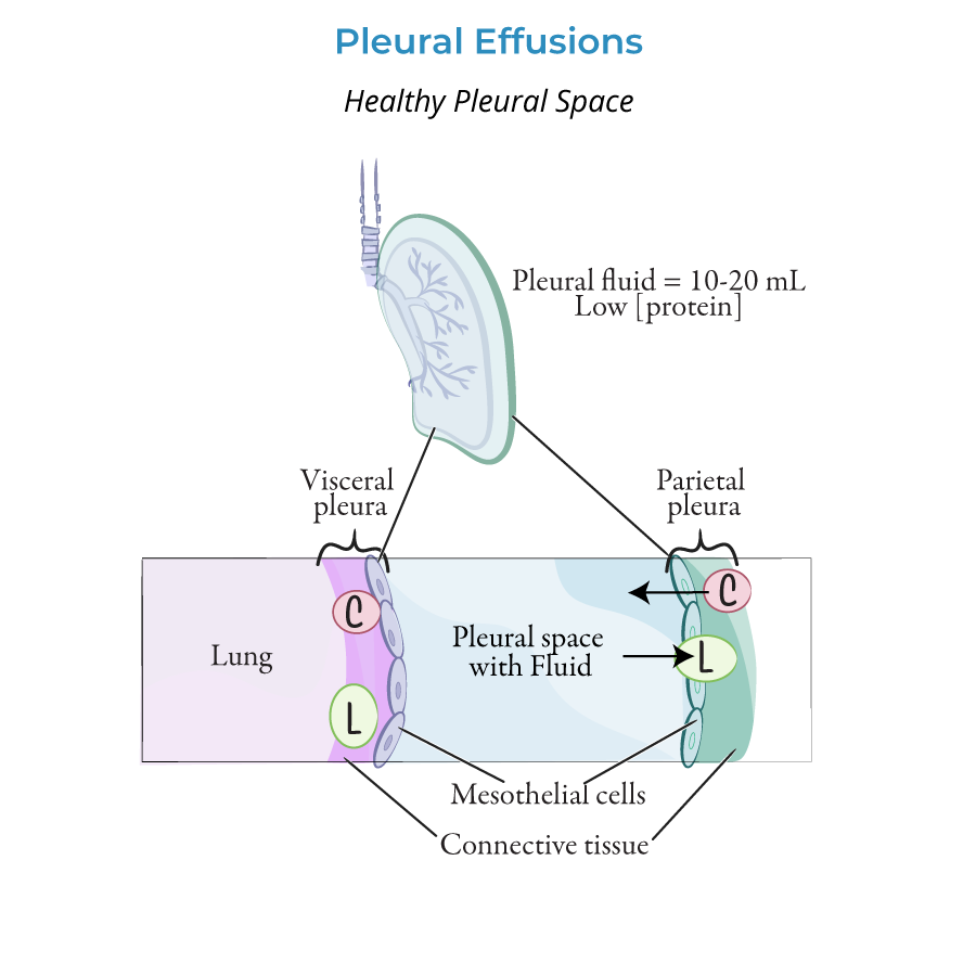
The pleural sac comprises the visceral pleura, which forms the outermost layer of the lungs, and the parietal pleura, which lines the pleural cavities in the thorax. The two pleura are continuous with one another at the hilum on the mediastinal surfaces of the lungs.
The thin space between the visceral and parietal pleura is called the pleural space; it contains a thin layer of pleural fluid.
The right lung is divided by the horizontal and oblique fissures to form the superior, middle, and inferior lobes.
The left lung is divided by the oblique fissure into superior and inferior lobes; the cardiac notch represents where the heart sits.
The lungs are further divided into bronchopulmonary segments, which we don't show here; the right lung has 10 segments and the left has 8-9 segments.
The pleural sac envelops the lungs; the pleural sac reduces friction and allows the lungs to move freely during ventilation.
The pleural sac comprises the visceral pleura, which forms the outermost layer of the lungs, and the parietal pleura, which lines the pleural cavities in the thorax. The two pleura are continuous with one another at the hilum on the mediastinal surfaces of the lungs.
The thin space between the visceral and parietal pleura is called the pleural space; it contains a thin layer of pleural fluid.
 Label two recesses where the lungs do not fill the pleural sac:
The costomediastinal recess is anterior, where the costal pleura is opposed to the mediastinal pleura. This recess is larger on the left side in the pleura that overlies the heart.
The costodiaphragmatic recess occurs in the inferior pleural cavities where the costal pleura and diaphragmatic pleura are opposed.
These recesses allow for additional lung expansion during the forced inspiration. They are also clinically important sites of fluid accumulation during pleural effusions, which can put pressure on the lungs and impair breathing.
Pleural Effusion
Pleurisy is inflammation of the pleura, and can lead to sharp chest pain upon breathing or coughing.
Pleurisy aka pleuritis can be caused by respiratory infection; it can also be caused by traumatic injury or pneumothorax.
Label two recesses where the lungs do not fill the pleural sac:
The costomediastinal recess is anterior, where the costal pleura is opposed to the mediastinal pleura. This recess is larger on the left side in the pleura that overlies the heart.
The costodiaphragmatic recess occurs in the inferior pleural cavities where the costal pleura and diaphragmatic pleura are opposed.
These recesses allow for additional lung expansion during the forced inspiration. They are also clinically important sites of fluid accumulation during pleural effusions, which can put pressure on the lungs and impair breathing.
Pleural Effusion
Pleurisy is inflammation of the pleura, and can lead to sharp chest pain upon breathing or coughing.
Pleurisy aka pleuritis can be caused by respiratory infection; it can also be caused by traumatic injury or pneumothorax.
Lungs
Anterior View
First we show the tracheobronchial tree.
Then, we draw the right and left lungs; notice that the left lung is smaller.
The top, aka apex of the lung is dome-like, and extends above the first rib and into the root of the neck.
The base of the lungs is wider and concave; because it rests on the thoracic diaphragm, the base is also called the diaphragmatic surface.
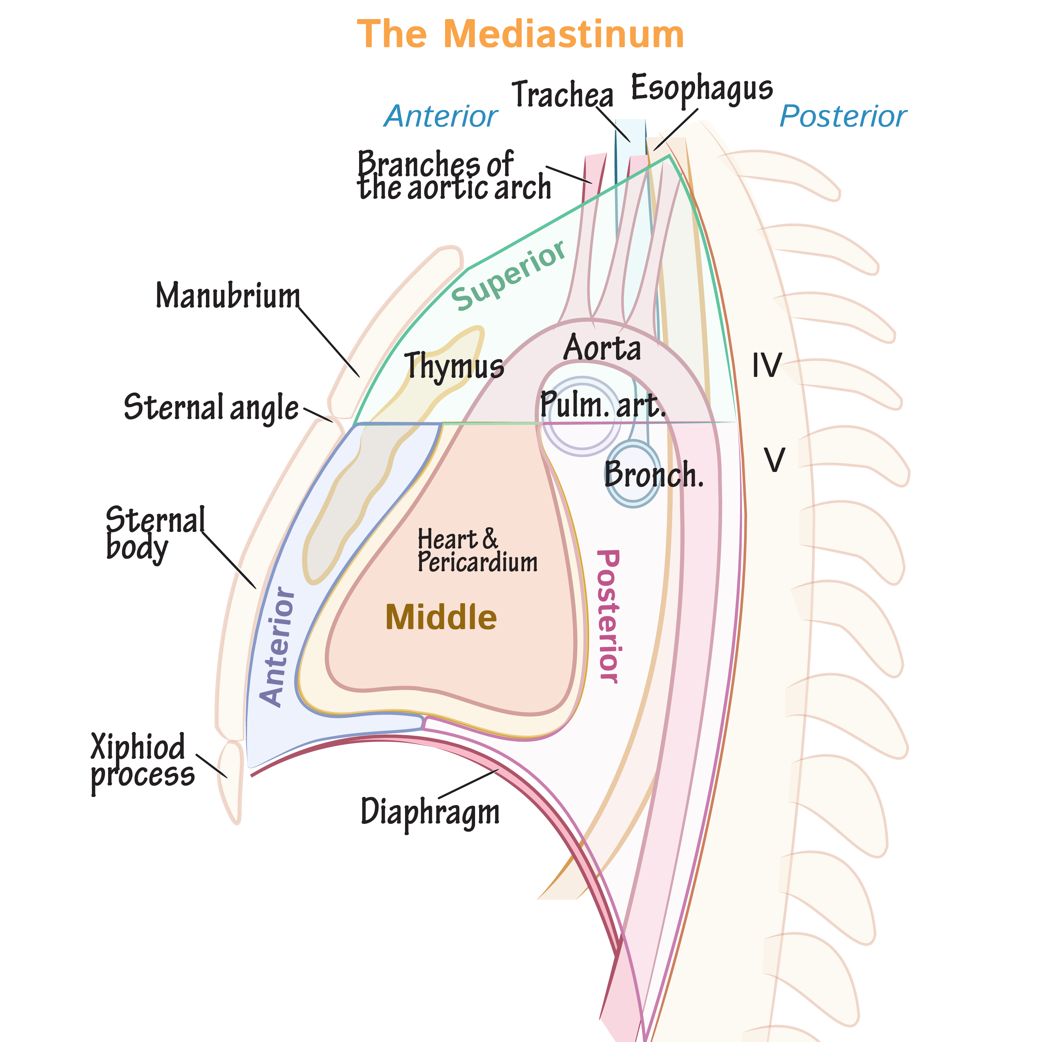
The costal surface of the lungs faces the ribs, and the mediastinal surface faces the mediastinum, which is the space between the lungs that houses the heart, esophagus, trachea, and neurovascular structures.
The right lung is divided by the horizontal and oblique fissures to form the superior, middle, and inferior lobes.
The left lung is divided by the oblique fissure into superior and inferior lobes; the cardiac notch represents where the heart sits.
The lungs are further divided into bronchopulmonary segments, which we don't show here; the right lung has 10 segments and the left has 8-9 segments.
Medial View
We re-draw the left lung and label its apex and base. We show the oblique fissure separating the superior and inferior lobes.
The hilum is where the root of the lung emerges from the lung.
The root comprises the bronchus and pulmonary arteries and veins; nervous and lymphatic structures also pass through here.
The pleural sleeve aka mesopneumonium, covers the structures of the root of the lung.
The pulmonary ligament where the mediastinal pleura folds upon itself at the inferior hilum.
Two key surface indentations:
Anterior to the hilum we see the cardiac impression.
Just above the hilum we see the groove for the arch of the aorta.
Pleural sac
Label two recesses where the lungs do not fill the pleural sac:
The costomediastinal recess is anterior, where the costal pleura is opposed to the mediastinal pleura. This recess is larger on the left side in the pleura that overlies the heart.
The costodiaphragmatic recess occurs in the inferior pleural cavities where the costal pleura and diaphragmatic pleura are opposed.
These recesses allow for additional lung expansion during the forced inspiration. They are also clinically important sites of fluid accumulation during pleural effusions, which can put pressure on the lungs and impair breathing.
Clinical Correlations
Pneumothorax
Occurs when a ruptured pleural sac allows air to leak into the chest cavity, which increases pressure on the lung leading to its collapse. Pneumothorax can be spontaneous or traumatic and requires immediate treatment.