

Start your One-Week Free Trial
Already subscribed? Log in »

Gastrointestinal Tract Tunics
Here we will learn the anatomy of the four layers, aka, tunics, of the gastrointestinal tract.
For additional histological details, see our full tutorial..
Let’s begin with a generalized cross section of the GI tract; then, we’ll learn about segmental differences along the tract.
From deep to superficial, show the following layers:
The mucosa lines the lumen of the GI tract; it comprises three sublayers:
The innermost layer is the epithelium, which comes is in direct contact with the contents of the GI tract.
The lamina propria is the middle layer, and it comprises loose connective tissue.
The muscularis mucosae is the outermost layer of the mucosa, and it comprises an inner circular and outer longitudinal layer.
The submucosa is next; notice that its inner surface follows the contours of the mucosa.
It houses the submucosal plexus, one of two special neural networks that regulate GI functioning, as well as vessels, glands, and lymphatic structures.
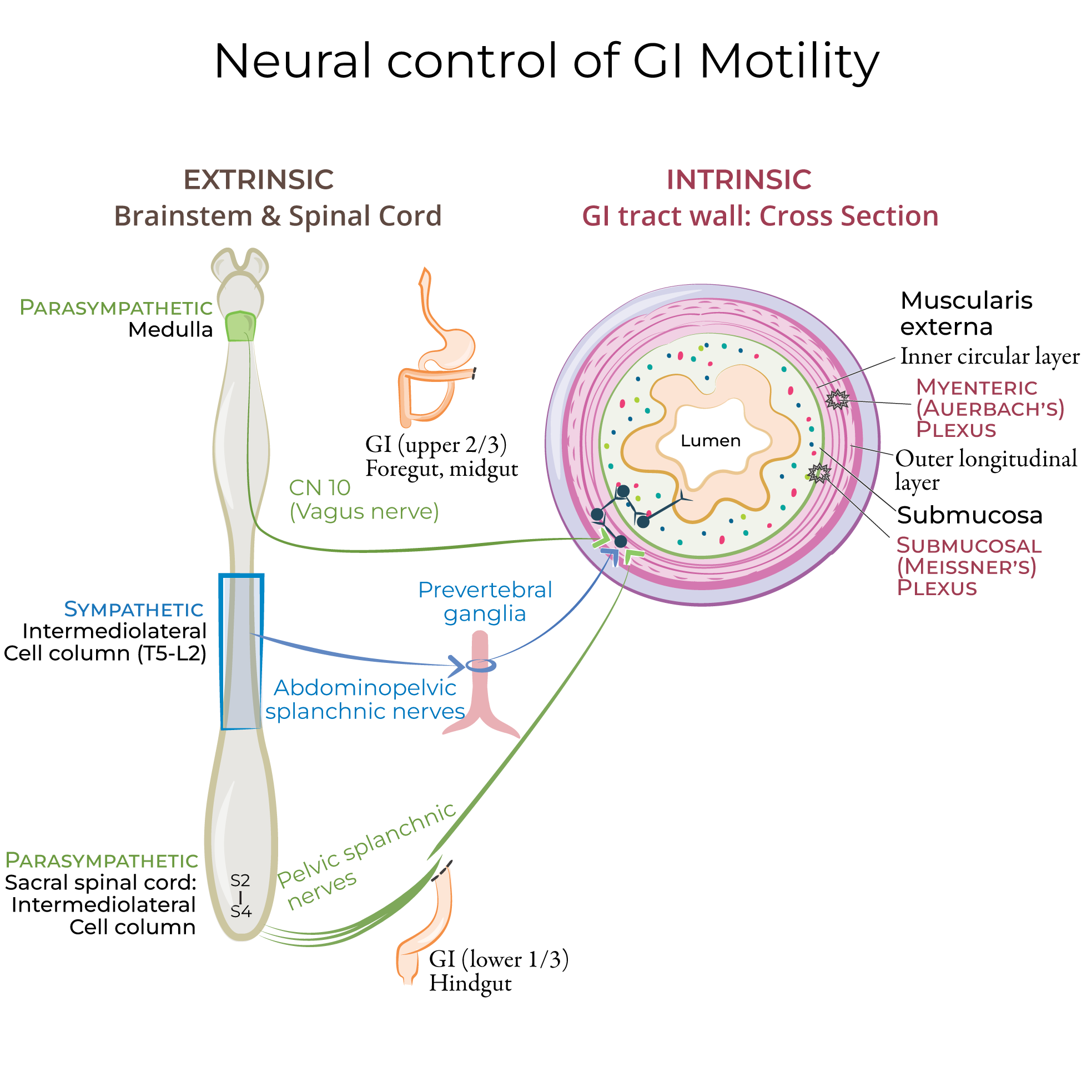 The muscularis externa comprises two sublayers:
An inner circular layer, which comprises muscle fibers that encircle the diameter of the GI tract, and,
An outer longitudinal layer, which comprises muscle fibers that run lengthwise along the GI tract.
Indicate that the myenteric nerve plexus lies between the inner and outer layers of the muscularis externa.
The muscularis externa is responsible for peristalsis, which propels foods and liquids through the GI tract.
Lastly, show that the outermost layer comprises either adventitia or serosa, depending on its position in the abdominal cavity.
As a general rule, serosa surrounds organs that are intraperitoneal; in other words, they are suspended in the abdomen by visceral peritoneum (such as the stomach).
Adventitia surrounds organs that are retroperitoneal; they are adhered to the abdominal wall (such as the ascending colon).
The muscularis externa comprises two sublayers:
An inner circular layer, which comprises muscle fibers that encircle the diameter of the GI tract, and,
An outer longitudinal layer, which comprises muscle fibers that run lengthwise along the GI tract.
Indicate that the myenteric nerve plexus lies between the inner and outer layers of the muscularis externa.
The muscularis externa is responsible for peristalsis, which propels foods and liquids through the GI tract.
Lastly, show that the outermost layer comprises either adventitia or serosa, depending on its position in the abdominal cavity.
As a general rule, serosa surrounds organs that are intraperitoneal; in other words, they are suspended in the abdomen by visceral peritoneum (such as the stomach).
Adventitia surrounds organs that are retroperitoneal; they are adhered to the abdominal wall (such as the ascending colon).
 The following GI segments are suspended from the abdominal walls and have serosa:
Stomach
Proximal duodenum
Jejunum and ileum
Cecum and appendix
Transverse colon
Sigmoid colon
The following organs are adhered to the abdominal wall and have adventitia on at least one surface:
Distal duodenum
The Pancreas
Ascending colon
Descending colon
Rectum (technically, the proximal portion has serosa, and only the distal part has adventitia).
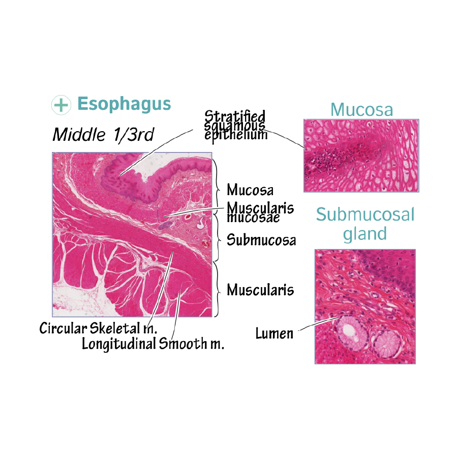
The esophageal mucosa comprises stratified squamous epithelia, which protects against abrasions from swallowed foods. Most of the GI tract mucosa is simple columnar epithelium.
The submucosa of the esophagus is rich in elastic fibers, to accommodate foods and liquids.
The following GI segments are suspended from the abdominal walls and have serosa:
Stomach
Proximal duodenum
Jejunum and ileum
Cecum and appendix
Transverse colon
Sigmoid colon
The following organs are adhered to the abdominal wall and have adventitia on at least one surface:
Distal duodenum
The Pancreas
Ascending colon
Descending colon
Rectum (technically, the proximal portion has serosa, and only the distal part has adventitia).
The esophageal mucosa comprises stratified squamous epithelia, which protects against abrasions from swallowed foods. Most of the GI tract mucosa is simple columnar epithelium.
The submucosa of the esophagus is rich in elastic fibers, to accommodate foods and liquids.
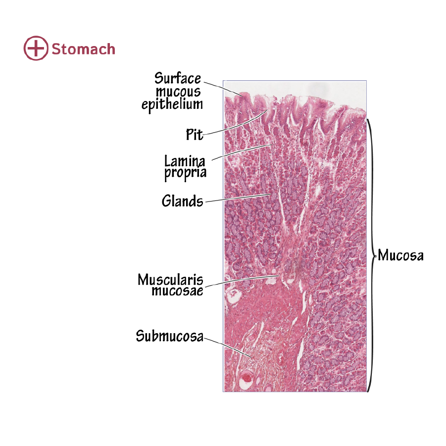
 The mucosa of the stomach houses gastric pits that open to gastric glands. The gastric glands secrete a variety of substances that aid in protection of the stomach lining and digestion (including mucus, gastric acids and hormones).
The muscularis externa layer of the stomach has a unique third sublayer of muscle fibers, called the oblique layer, which wraps obliquely around the stomach to enhance mixing and churning of foods.
The mucosa of the stomach houses gastric pits that open to gastric glands. The gastric glands secrete a variety of substances that aid in protection of the stomach lining and digestion (including mucus, gastric acids and hormones).
The muscularis externa layer of the stomach has a unique third sublayer of muscle fibers, called the oblique layer, which wraps obliquely around the stomach to enhance mixing and churning of foods.
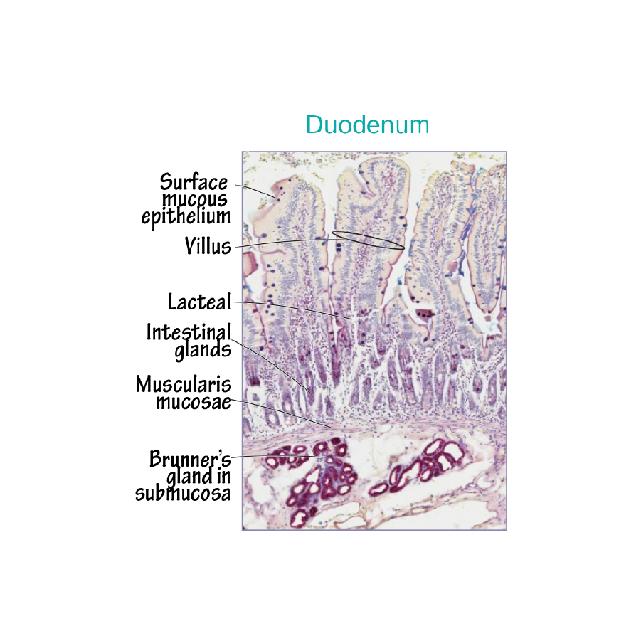
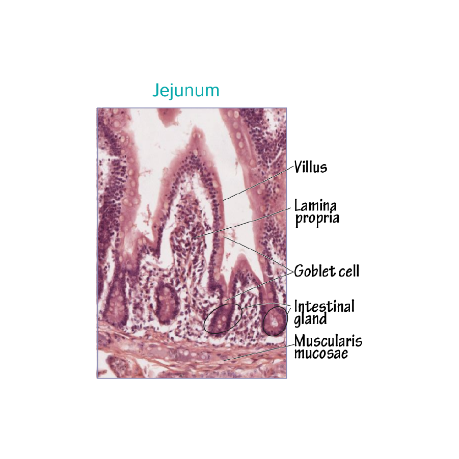
 The mucosa of the small intestine is arranged in circular folds (aka, plicae circulares, aka, valves of Kerckring), which are covered with villi. Circular folds are most prominent within the jejunum.
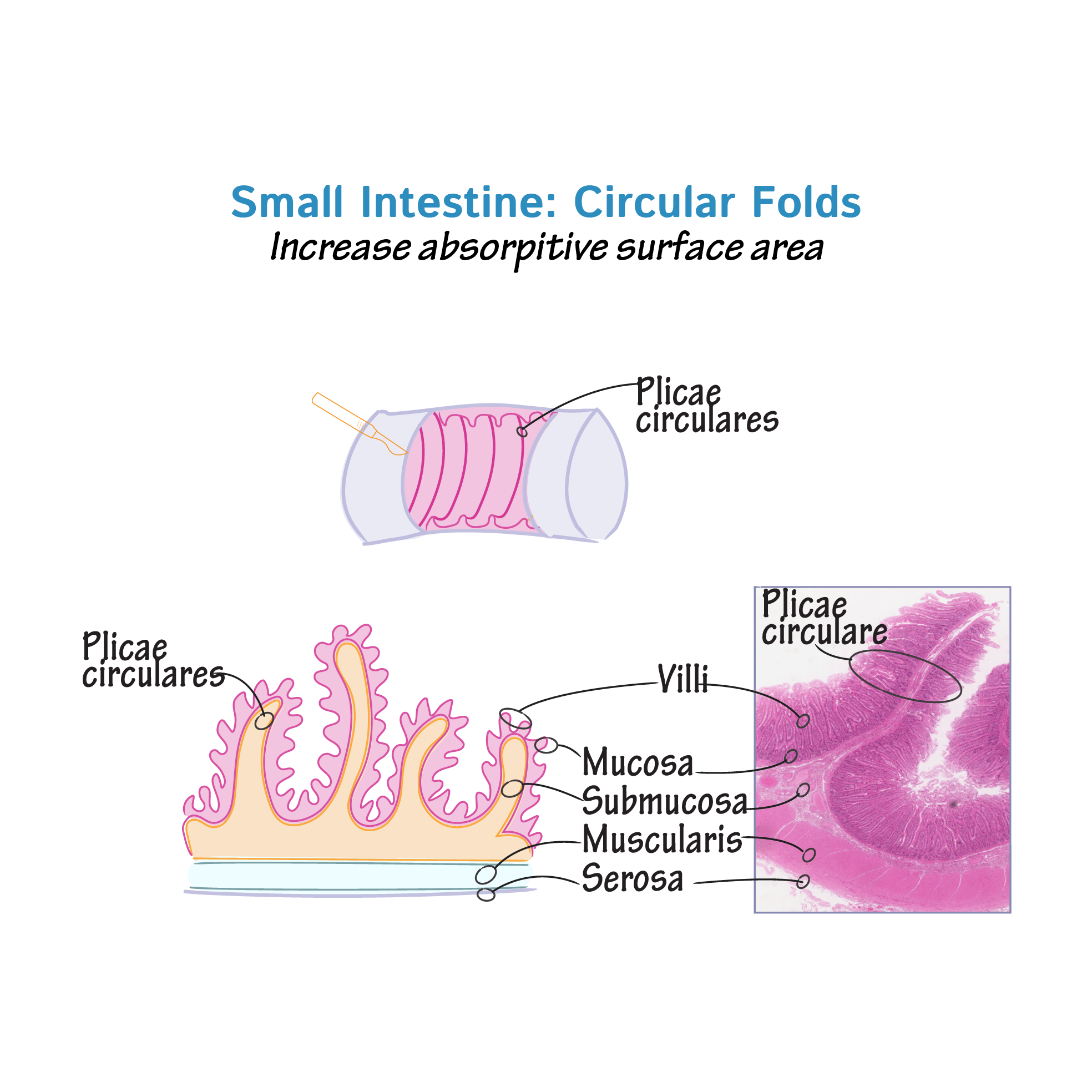 Circular folds increase the surface area of the small intestine, and, therefore, the amount of nutrient absorption. The small intestine also houses intestinal glands and goblet cells that aid in nutrient absorption and intestinal lubrication.
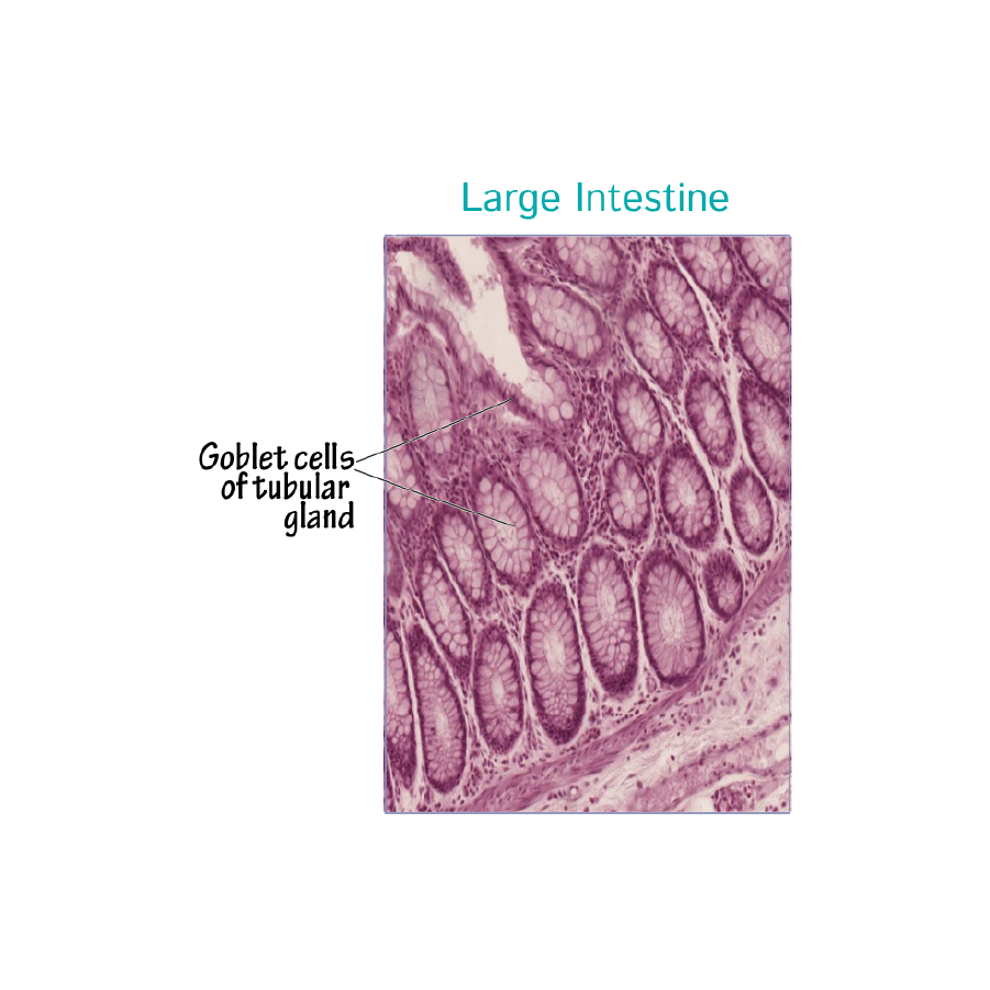
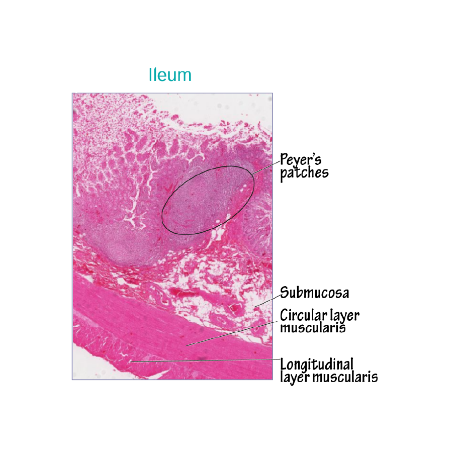 The mucosa of the large intestine houses intestinal glands and goblet cells, as well as lymphatic structures (as we learn elsewhere, lymphatic structures are also present in the submucosa of the large intestine).
The muscularis externa layer of the large intestine is also unique; the outer longitudinal layer forms three distinct, visible bands called taeniae coli that run lengthwise along most of the large intestine (at the rectum, the separate bands converge to form a single layer of muscle).
We can see in our diagram that the taeniae coli “bunch” the colon into pouches called haustra. Contractions of the taeniae coli shorten the length of the colon and propel fecal matter through it.
The mucosa of the large intestine houses intestinal glands and goblet cells, as well as lymphatic structures (as we learn elsewhere, lymphatic structures are also present in the submucosa of the large intestine).
The muscularis externa layer of the large intestine is also unique; the outer longitudinal layer forms three distinct, visible bands called taeniae coli that run lengthwise along most of the large intestine (at the rectum, the separate bands converge to form a single layer of muscle).
We can see in our diagram that the taeniae coli “bunch” the colon into pouches called haustra. Contractions of the taeniae coli shorten the length of the colon and propel fecal matter through it.
Cross Section of the GI tract
The muscularis externa comprises two sublayers:
An inner circular layer, which comprises muscle fibers that encircle the diameter of the GI tract, and,
An outer longitudinal layer, which comprises muscle fibers that run lengthwise along the GI tract.
Indicate that the myenteric nerve plexus lies between the inner and outer layers of the muscularis externa.
The muscularis externa is responsible for peristalsis, which propels foods and liquids through the GI tract.
Lastly, show that the outermost layer comprises either adventitia or serosa, depending on its position in the abdominal cavity.
As a general rule, serosa surrounds organs that are intraperitoneal; in other words, they are suspended in the abdomen by visceral peritoneum (such as the stomach).
Adventitia surrounds organs that are retroperitoneal; they are adhered to the abdominal wall (such as the ascending colon).
The following GI segments are suspended from the abdominal walls and have serosa:
Stomach
Proximal duodenum
Jejunum and ileum
Cecum and appendix
Transverse colon
Sigmoid colon
The following organs are adhered to the abdominal wall and have adventitia on at least one surface:
Distal duodenum
The Pancreas
Ascending colon
Descending colon
Rectum (technically, the proximal portion has serosa, and only the distal part has adventitia).
Dey differences along the GI tract
The mucosa of the stomach houses gastric pits that open to gastric glands. The gastric glands secrete a variety of substances that aid in protection of the stomach lining and digestion (including mucus, gastric acids and hormones).
The muscularis externa layer of the stomach has a unique third sublayer of muscle fibers, called the oblique layer, which wraps obliquely around the stomach to enhance mixing and churning of foods.
The mucosa of the small intestine is arranged in circular folds (aka, plicae circulares, aka, valves of Kerckring), which are covered with villi. Circular folds are most prominent within the jejunum.
Circular folds increase the surface area of the small intestine, and, therefore, the amount of nutrient absorption. The small intestine also houses intestinal glands and goblet cells that aid in nutrient absorption and intestinal lubrication.
The mucosa of the large intestine houses intestinal glands and goblet cells, as well as lymphatic structures (as we learn elsewhere, lymphatic structures are also present in the submucosa of the large intestine).
The muscularis externa layer of the large intestine is also unique; the outer longitudinal layer forms three distinct, visible bands called taeniae coli that run lengthwise along most of the large intestine (at the rectum, the separate bands converge to form a single layer of muscle).
We can see in our diagram that the taeniae coli “bunch” the colon into pouches called haustra. Contractions of the taeniae coli shorten the length of the colon and propel fecal matter through it.