

Start your One-Week Free Trial
Already subscribed? Log in »

Anterior Thigh - Neurovascular Structures
Here we'll learn key vessels and nerves in the anterior thigh.
Anterior Thigh Muscles
!Anterior thigh muscles
Medial Thigh Muscles
!Medial thigh muscles
 These vessels enter the femoral triangle close to the femoral nerve, at the middle of the femoral triangle, but notice that the nerve is not enclosed in the femoral sheath.
The femoral artery and vein travel in the adductor canal with the saphenous nerve.
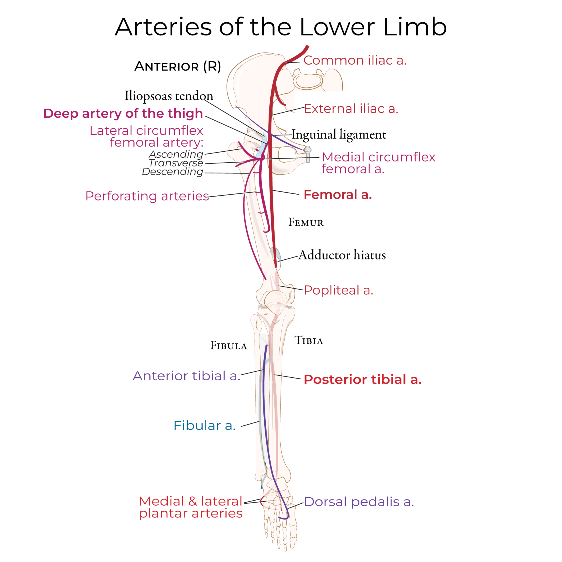
The femoral artery gives rise to the deep artery of the thigh (aka, profunda femoris artery); this vessel dives deep to serve muscles in the medial and posterior compartments.
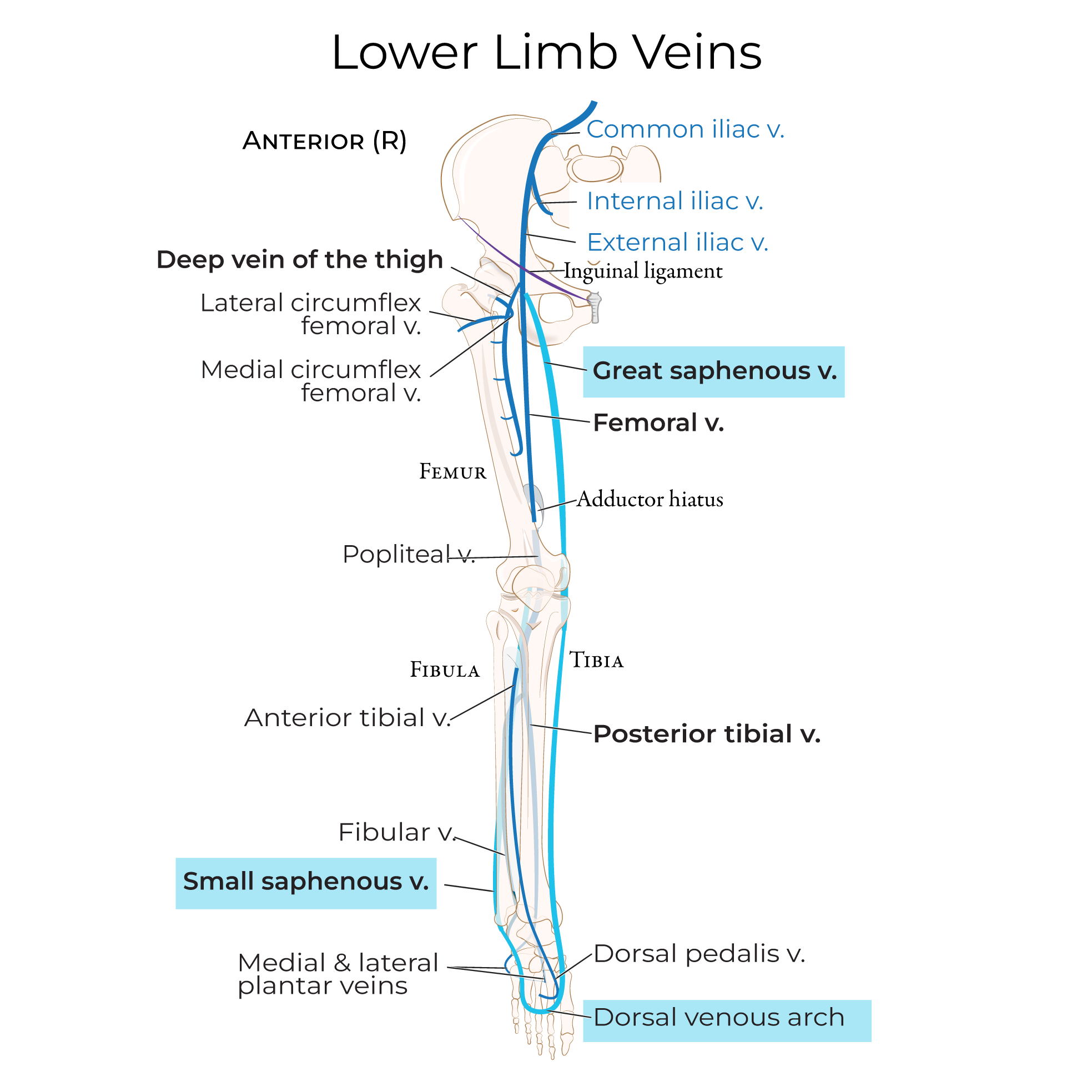
The great saphenous vein drains into the femoral vein within the femoral triangle; the great saphenous vein is a subcutaneous vessel that originates in the foot and travels superiorly along the medial side of the lower extremity.
These vessels enter the femoral triangle close to the femoral nerve, at the middle of the femoral triangle, but notice that the nerve is not enclosed in the femoral sheath.
The femoral artery and vein travel in the adductor canal with the saphenous nerve.
The femoral artery gives rise to the deep artery of the thigh (aka, profunda femoris artery); this vessel dives deep to serve muscles in the medial and posterior compartments.
The great saphenous vein drains into the femoral vein within the femoral triangle; the great saphenous vein is a subcutaneous vessel that originates in the foot and travels superiorly along the medial side of the lower extremity.
 Distally, the vessels pass under the anteromedial intermuscular septum, then travel posteriorly through the adductor hiatus to the back of the knee.
 See more
Review Flashcards
Superficial Thigh
Muscles:
- Iliopsoas (the combined fibers of iliacus and psoas major) crosses the hip joint and the nearby pectineus.
- Quadriceps femoris group makes up the bulk of the anterior thigh.
- Tensor fascia latae runs laterally along the thigh.
- Adductor longus, adductor magnus, and gracilis comprise the medial compartment (adductor brevis is too deep to see here).
- Sartorius is mostly "cut out" so we can see the neurovascular structures in the adductor canal, which is deep to sartorius.
Adductor canal
- Aka, the subsartorial canal, is a long, narrow passageway that arises at about the midpoint of the anterior thigh between qudriceps femoris and the adductors. This space provides a protected groove for neurovascular structures.
Femoral Triangle
Bound by:
The inguinal ligament, superiorly
Adductor longus, medially
And sartorius, laterally (we've removed sartorius)
The apex of the femoral triangle is where sartorius crosses over the adductor longus.
The femoral triangle demarcates the area where key nerves and vessels pass between the pelvis and the thigh; it's a helpful landmark during dissection.
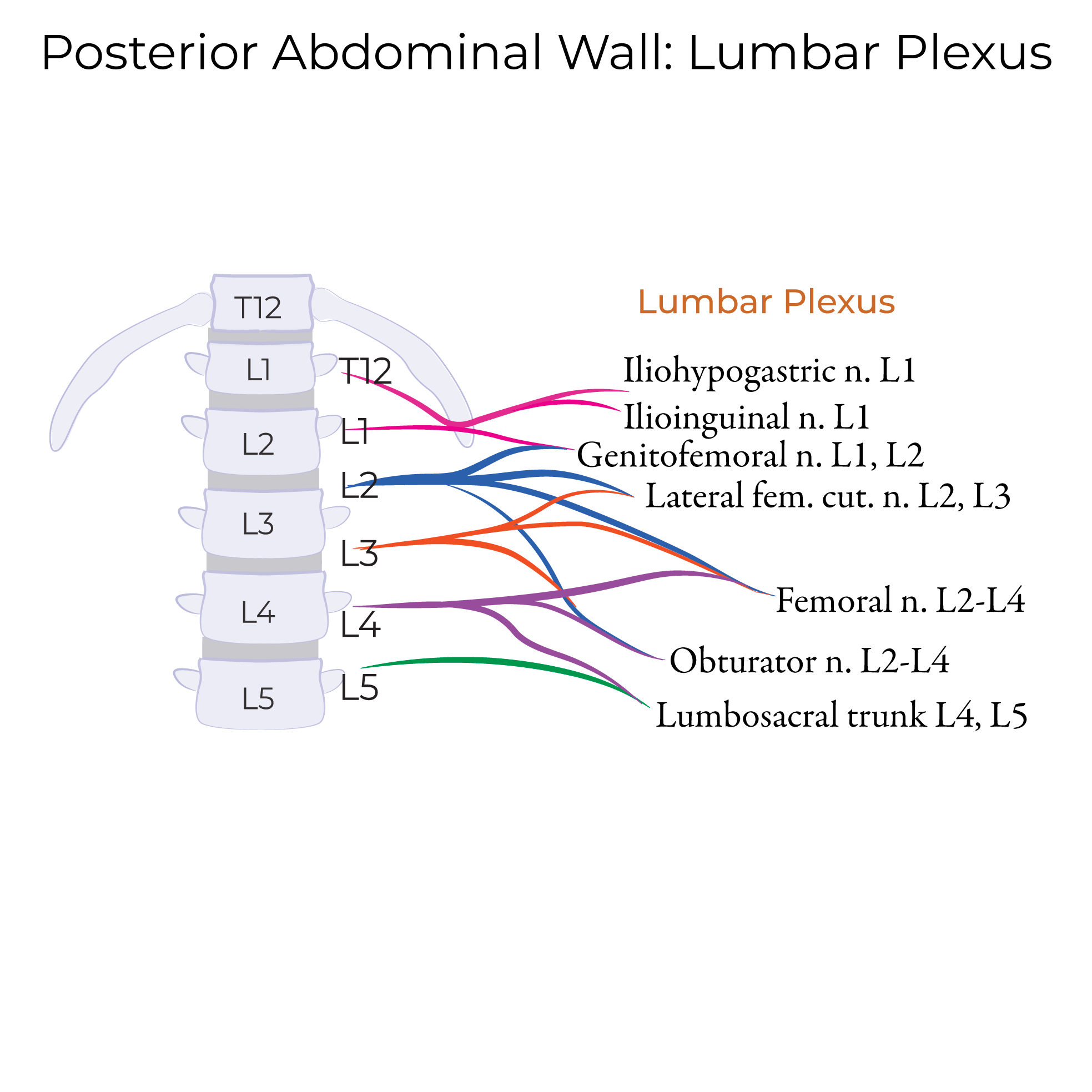
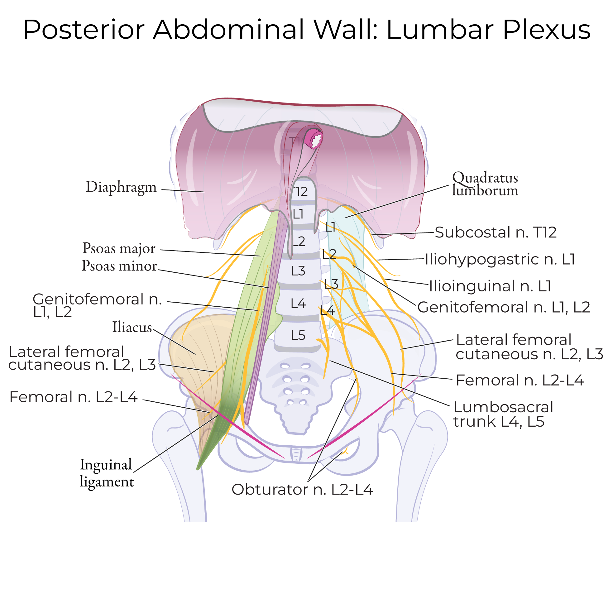
Anterior Thigh Inner - Lumbar Plexus
- The lateral femoral cutaneous nerve (L2 & L3) passes under the inguinal ligament; it provides sensation to the anterior and lateral thigh.
- The femoral nerve (L2-L4) passes under the inguinal it gives rise to several terminal branches that serve the skin and muscles of the anterior thigh.
Femoral sheath
A 3-4 inch fascial tube; it is a continuation of abdominal fascial layers (the transveralis abdominis and iliopsoas fascia, specifically).
The femoral sheath provides passage for an artery, vein, and lymph nodes to pass from the pelvis to the thigh; the sheath is subdivided to create specific fascial channels for each structure.
The external iliac artery and vein become the femoral artery and vein when they pass deep to the inguinal ligament.
These vessels enter the femoral triangle close to the femoral nerve, at the middle of the femoral triangle, but notice that the nerve is not enclosed in the femoral sheath.
The femoral artery and vein travel in the adductor canal with the saphenous nerve.
The femoral artery gives rise to the deep artery of the thigh (aka, profunda femoris artery); this vessel dives deep to serve muscles in the medial and posterior compartments.
The great saphenous vein drains into the femoral vein within the femoral triangle; the great saphenous vein is a subcutaneous vessel that originates in the foot and travels superiorly along the medial side of the lower extremity.
Distally, the vessels pass under the anteromedial intermuscular septum, then travel posteriorly through the adductor hiatus to the back of the knee.
Deeper Thigh
Muscles
- Obturator externus and iliopsoas.
- Three of the four quadriceps femoris muscles: vastus intermedius, vastus lateralis, and vastus medialis. Note that rectus femoris is omitted.
- Portion of pectineus that emerges from under the adductors, but "cut" it before it crosses obturator externus.
- Adductor brevis, adductor magnus, and gracilis; notice we've left out the more superficial adductor longus.
- Note that we've omitted sartorius and tensor fascia latae.
Neurovasculature
- The femoral nerve: we show its muscular branches in the anterior thigh and the saphenous nerve in the adductor canal.
- The external iliac artery and vein becoming the femoral artery and vein (we've omitted the femoral sheath for simplicity on this diagram). We "cut out" the middle portion of these vessels, then show where they pass under the anteromedial intermuscular septum and through the adductor hiatus distally.
- Deep artery of the thigh as it travels slightly laterally; indicate that it gives rise to:
- Anterior and posterior branches of the obturator nerve (L2-L4), which are separated by adductor brevis.
Summary of Femoral Artery & Branches
- Deep femoral artery/profunda femoris artery: Splits off femoral artery within 5 cm from the inguinal ligament, passes between pectineus and adductor longus and gives rise to 3-4 perforating branches that pass through the adductors. Supplies medial, posterior, and lateral aspect of the anterior compartment.
- Medial circumflex femoral artery: Arises from deep femoral artery (sometimes from femoral artery). Passes medially/posteriorly to enter gluteal region. Gives rise to posterior retinacular arteries, transverse and ascending arteries. Supplies femoral head and neck.
- Lateral circumflex artery: Arises from deep femoral artery (sometimes from femoral artery). Passes laterally under sartorius and rectus femoris, then divides into ascending, transverse, and descending arteries. Ascending supplies gluteal region; transverse passes around femur; descending joins genicular peri-articular anastomoses.
- Superficial epigastric artery: Arises from the femoral artery near to the inguinal ligament and travels superiorly towards the umbilicus; it is a relatively superficial vessel that supplies the skin.
- Superficial circumflex iliac artery: Arises from femoral artery, near superficial epigastric artery; travels towards anterior superior iliac spine and supplies the skin.
- Superficial and Deep external pudendal arteries: Arises from femoral artery and travels towards external genitalia to supply the skin.
- Descending genicular artery: Arises from femoral artery and travels to the medial knee where it anastomoses with the medial superior genicular artery. Supplies vastus medialis, adductor magnus, and medial skin of the thigh.
Summary of Anterior Thigh Innervation
- Femoral nerve: L2-L4, largest branch of lumbar plexus, arises within psoas major and enters thigh in femoral triangle deep to the inguinal canal, where it gives rise to several motor branches that serve anterior thigh muscles.
- Obturator nerve: also arises from the lumbar plexus (L2-4), supplies the obturator externus, pectineus, and muscles of the medial thigh; it also serves the skin of the medial thigh.
- Lateral femoral cutaneous (L2, L3) nerve supplies the anterior and lateral thigh.
- Genitofemoral nerve (L1, L2) serves the skin of the upper anterior thigh.
- Ilioinguinal nerve (L1) serves the skin of the upper medial thigh.
See more