

Start your One-Week Free Trial
Already subscribed? Log in »

Veins - Lower Extremity
Here we will learn the deep and superficial veins of the lower extremity. Be aware that there is a great deal of variation in venous patterns, and that anastomoses are quite common.
The major superficial veins include the great and small saphenous veins; these are located in the subcutaneous tissues.
The deep veins travel with the arteries, and share their names; they are often paired (though we’ll draw them as single vessels for simplicity). The deep veins run deep to the deep fascia of the lower limb.
 Perforating veins send blood from the superficial veins to the deep veins.
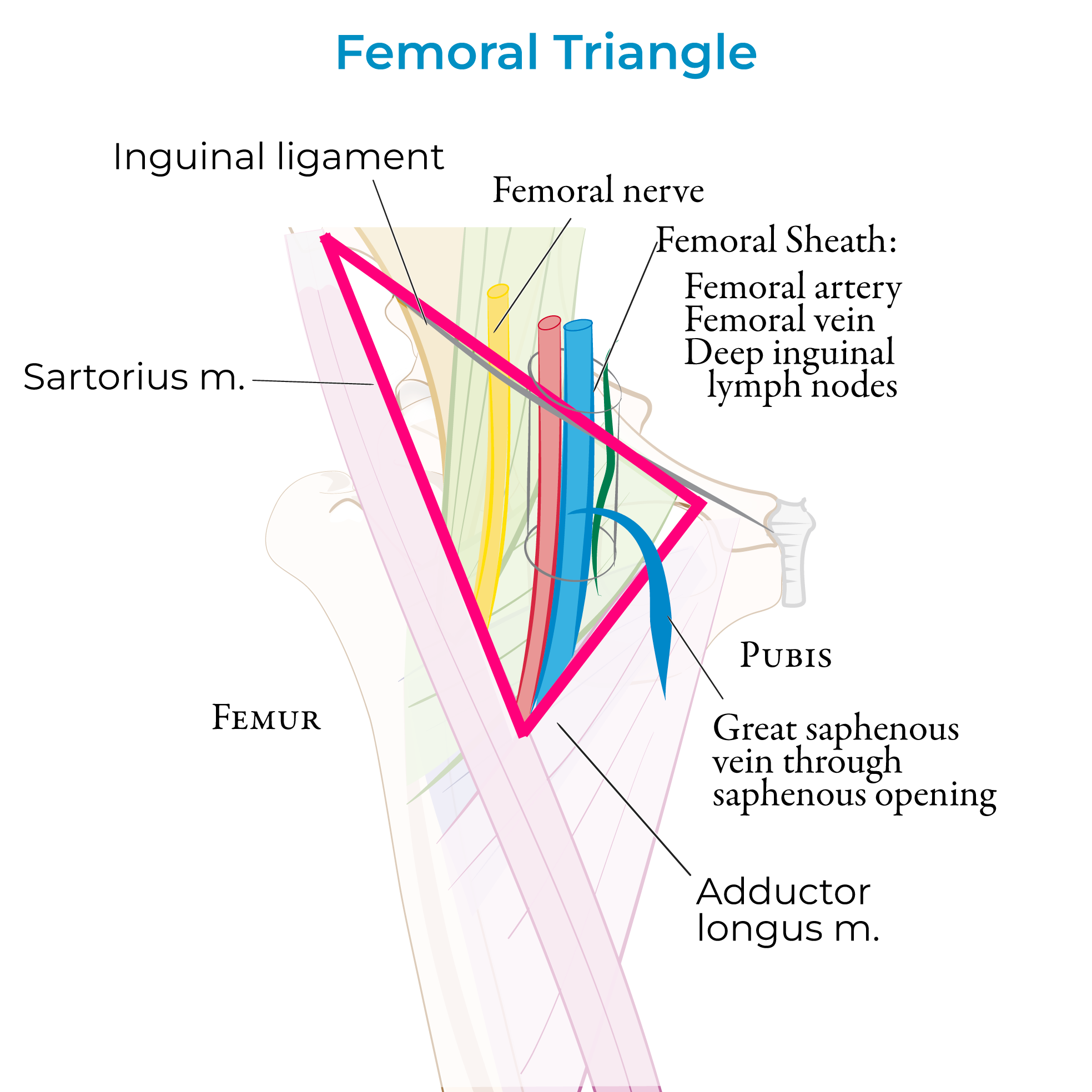
The femoral blood and lymph vessels and the nerves pass through the femoral triangle in a particular order from lateral to medial, which can be remembered with the acronym NAVL: Femoral Nerve, Artery, Vein, and Lymph.
Review the femoral triangle and neurovascular structures of the anterior thigh.
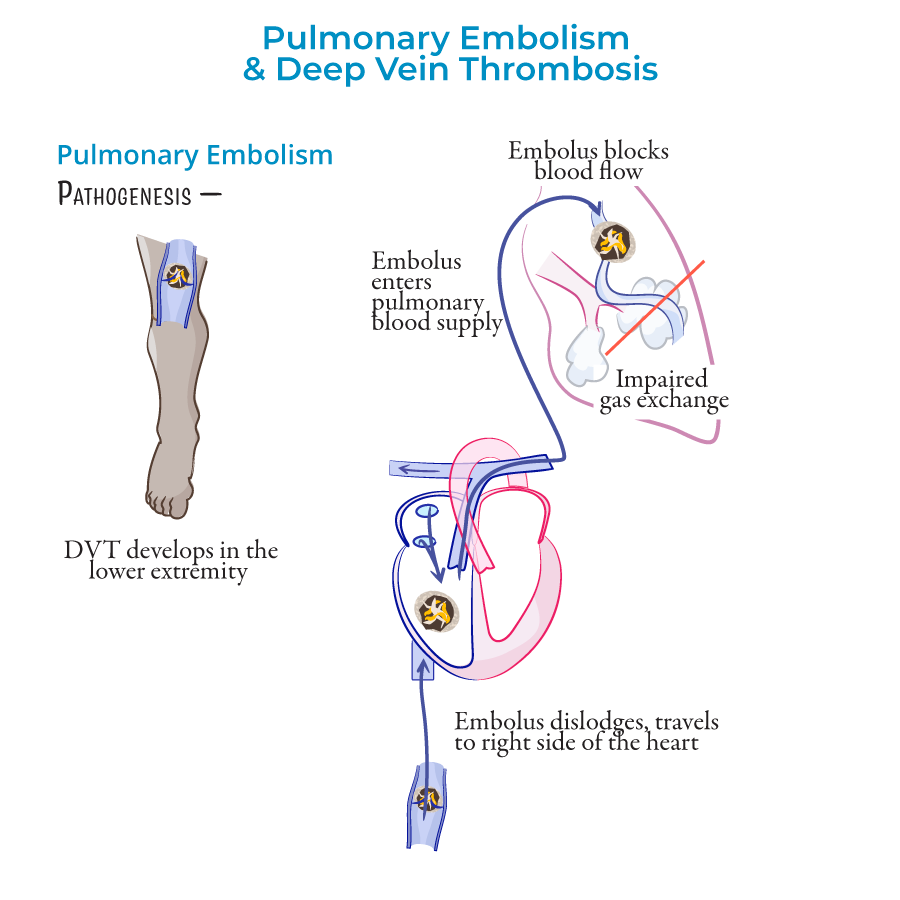
Deep vein thrombosis can be fatal when pieces of a clot in the lower limb break free and travel to the pulmonary artery, thereby obstructing blood flow through the lungs. We have a tutorial on this topic, if you’re interested, see our notes for the link.
Perforating veins send blood from the superficial veins to the deep veins.
The femoral blood and lymph vessels and the nerves pass through the femoral triangle in a particular order from lateral to medial, which can be remembered with the acronym NAVL: Femoral Nerve, Artery, Vein, and Lymph.
Review the femoral triangle and neurovascular structures of the anterior thigh.
Deep vein thrombosis can be fatal when pieces of a clot in the lower limb break free and travel to the pulmonary artery, thereby obstructing blood flow through the lungs. We have a tutorial on this topic, if you’re interested, see our notes for the link.
 We’ll draw the deep veins, first, since they receive blood from the superficial veins.
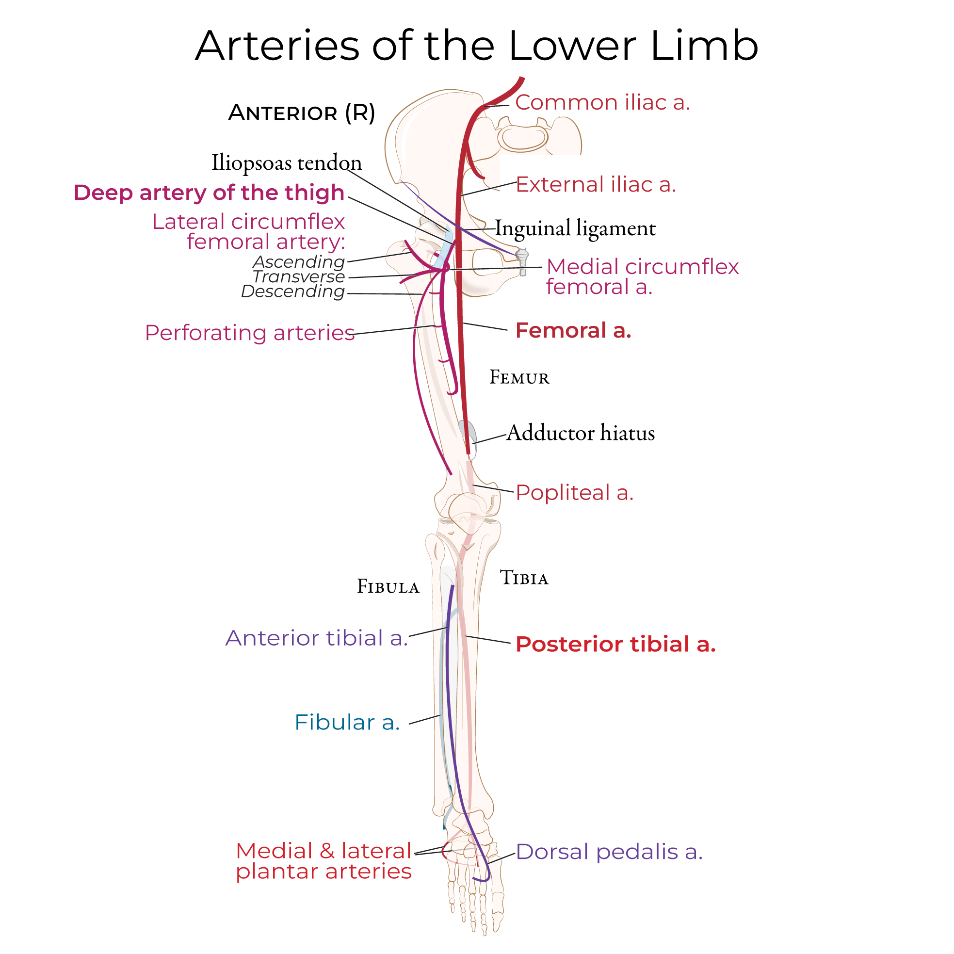
In the pelvis, the external and internal iliac veins merge to form the common iliac vein, which drains into the inferior vena cava.
From here we’ll trace venous return from the foot to the external iliac vein.
In the foot, the medial and lateral plantar veins drain into the posterior tibial and fibular veins; perforating veins from the dorsal venous arch give rise to the anterior tibial vein.
The three veins, posterior tibial, anterior tibial, and fibular, ascend through the leg and drain into the popliteal vein, which becomes the femoral vein.
The femoral vein ascends through the thigh and passes deep to the inguinal ligament to become the external iliac vein.
We’ll draw the deep veins, first, since they receive blood from the superficial veins.
In the pelvis, the external and internal iliac veins merge to form the common iliac vein, which drains into the inferior vena cava.
From here we’ll trace venous return from the foot to the external iliac vein.
In the foot, the medial and lateral plantar veins drain into the posterior tibial and fibular veins; perforating veins from the dorsal venous arch give rise to the anterior tibial vein.
The three veins, posterior tibial, anterior tibial, and fibular, ascend through the leg and drain into the popliteal vein, which becomes the femoral vein.
The femoral vein ascends through the thigh and passes deep to the inguinal ligament to become the external iliac vein.
 Perforating veins, as well as the medial and lateral circumflex veins, drain into the deep vein of the thigh, which drains into the femoral vein.
In the foot, the dorsal venous arch drains medially into the great saphenous vein, which passes behind the medial femoral condyle on its way to the proximal thigh, where it pierces the deep fascia and empties into the femoral vein. Note that, in the distal leg, the great saphenous vein travels with the saphenous nerve.
The great saphenous vein, itself, receives blood from the lateral and anterior cutaneous, superficial circumflex iliac, superficial epigastric, and external pudendal veins.
Laterally in the foot, the dorsal venous arch drains into the short saphenous vein, which travels posterior to the lateral malleolus and ascends through the posterior leg to the popliteal fossa, where it pierces the deep fascia and empties into the popliteal vein. Note that the short saphenous vein travels near the sural nerve in the distal leg.
Now, let’s show the leg in posterior view to get a better look at the deep and superficial veins.
Review muscles
Starting in the foot, the posterior tibial, anterior tibial, and fibular veins ascending through the leg; in the popliteal fossa, they combine to form the popliteal vein. The popliteal vein travels through the adductor hiatus and becomes the femoral vein.
Now, indicate the deep fascia of the lower limb; in the foot, show the superficial veins of the plantar venous network.
The small saphenous vein as it passes posteriorly around the lateral malleolus and travels over the deep fascia towards the popliteal fossa.
Behind the knee, the small saphenous vein pierces the deep fascia and passes between the two heads of gastrocnemius to empty into the popliteal vein.
Perforating veins, as well as the medial and lateral circumflex veins, drain into the deep vein of the thigh, which drains into the femoral vein.
In the foot, the dorsal venous arch drains medially into the great saphenous vein, which passes behind the medial femoral condyle on its way to the proximal thigh, where it pierces the deep fascia and empties into the femoral vein. Note that, in the distal leg, the great saphenous vein travels with the saphenous nerve.
The great saphenous vein, itself, receives blood from the lateral and anterior cutaneous, superficial circumflex iliac, superficial epigastric, and external pudendal veins.
Laterally in the foot, the dorsal venous arch drains into the short saphenous vein, which travels posterior to the lateral malleolus and ascends through the posterior leg to the popliteal fossa, where it pierces the deep fascia and empties into the popliteal vein. Note that the short saphenous vein travels near the sural nerve in the distal leg.
Now, let’s show the leg in posterior view to get a better look at the deep and superficial veins.
Review muscles
Starting in the foot, the posterior tibial, anterior tibial, and fibular veins ascending through the leg; in the popliteal fossa, they combine to form the popliteal vein. The popliteal vein travels through the adductor hiatus and becomes the femoral vein.
Now, indicate the deep fascia of the lower limb; in the foot, show the superficial veins of the plantar venous network.
The small saphenous vein as it passes posteriorly around the lateral malleolus and travels over the deep fascia towards the popliteal fossa.
Behind the knee, the small saphenous vein pierces the deep fascia and passes between the two heads of gastrocnemius to empty into the popliteal vein.
Key Points
Perforating veins send blood from the superficial veins to the deep veins.
The femoral blood and lymph vessels and the nerves pass through the femoral triangle in a particular order from lateral to medial, which can be remembered with the acronym NAVL: Femoral Nerve, Artery, Vein, and Lymph.
Review the femoral triangle and neurovascular structures of the anterior thigh.
Deep vein thrombosis can be fatal when pieces of a clot in the lower limb break free and travel to the pulmonary artery, thereby obstructing blood flow through the lungs. We have a tutorial on this topic, if you’re interested, see our notes for the link.
Anterior Diagram
Perforating veins, as well as the medial and lateral circumflex veins, drain into the deep vein of the thigh, which drains into the femoral vein.
In the foot, the dorsal venous arch drains medially into the great saphenous vein, which passes behind the medial femoral condyle on its way to the proximal thigh, where it pierces the deep fascia and empties into the femoral vein. Note that, in the distal leg, the great saphenous vein travels with the saphenous nerve.
The great saphenous vein, itself, receives blood from the lateral and anterior cutaneous, superficial circumflex iliac, superficial epigastric, and external pudendal veins.
Laterally in the foot, the dorsal venous arch drains into the short saphenous vein, which travels posterior to the lateral malleolus and ascends through the posterior leg to the popliteal fossa, where it pierces the deep fascia and empties into the popliteal vein. Note that the short saphenous vein travels near the sural nerve in the distal leg.
Posterior Leg