

Start your One-Week Free Trial
Already subscribed? Log in »


Pharyngeal Mucosa
Here we’ll learn about the mucosa of the pharynx, which is a common pathway for foods, liquids, and air.
 Now, show the thyroid cartilage, epiglottis, and the internal surface of the larynx; highlight the laryngeal inlet. Show the connective tissues between the hyoid bone and the thyroid cartilage and epiglottis.
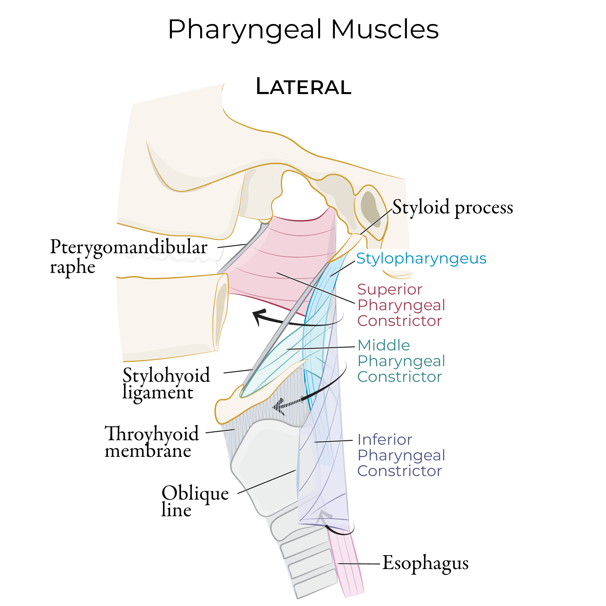
Indicate the pharyngeal constrictors, which form the half-cylinder muscular tube of the pharynx: label them as superior, middle, and inferior pharyngeal constrictors.
Now, show the thyroid cartilage, epiglottis, and the internal surface of the larynx; highlight the laryngeal inlet. Show the connective tissues between the hyoid bone and the thyroid cartilage and epiglottis.
Indicate the pharyngeal constrictors, which form the half-cylinder muscular tube of the pharynx: label them as superior, middle, and inferior pharyngeal constrictors.
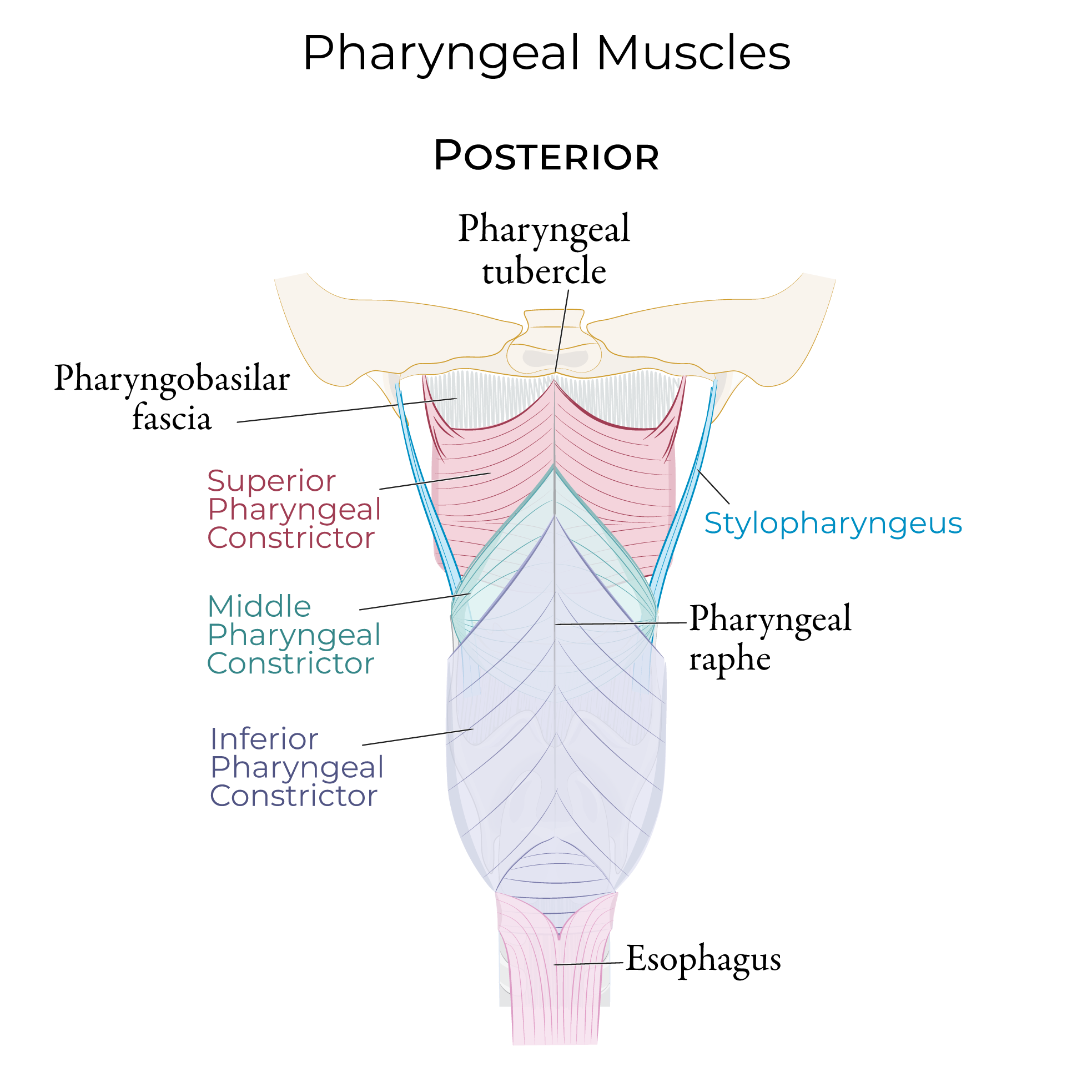 Show that the internal surface of the constrictors is covered and supported by the pharyngobasilar fascia; the external surface of the constrictors is covered by the thinner buccopharyngeal fascia.
 Now let’s learn the pharyngeal mucosa from posterior view, as if we’ve dissected down the midline of the throat.
Lateral View
Set up the diagram.
First, draw some key bony and cartilaginous features: the base of the skull, hyoid, and vertebral column.
Show the maxilla, which gives rise to the alveolar process with the upper teeth, and the palatine process, which forms the hard palate.
Show the soft palate extending posteriorly from the hard palate; the soft palate comprises the palatine aponeurosis and muscles. Indicate the skin over the anterior face.
Now, show that the hard and soft palates separate the nasal and oral cavities.
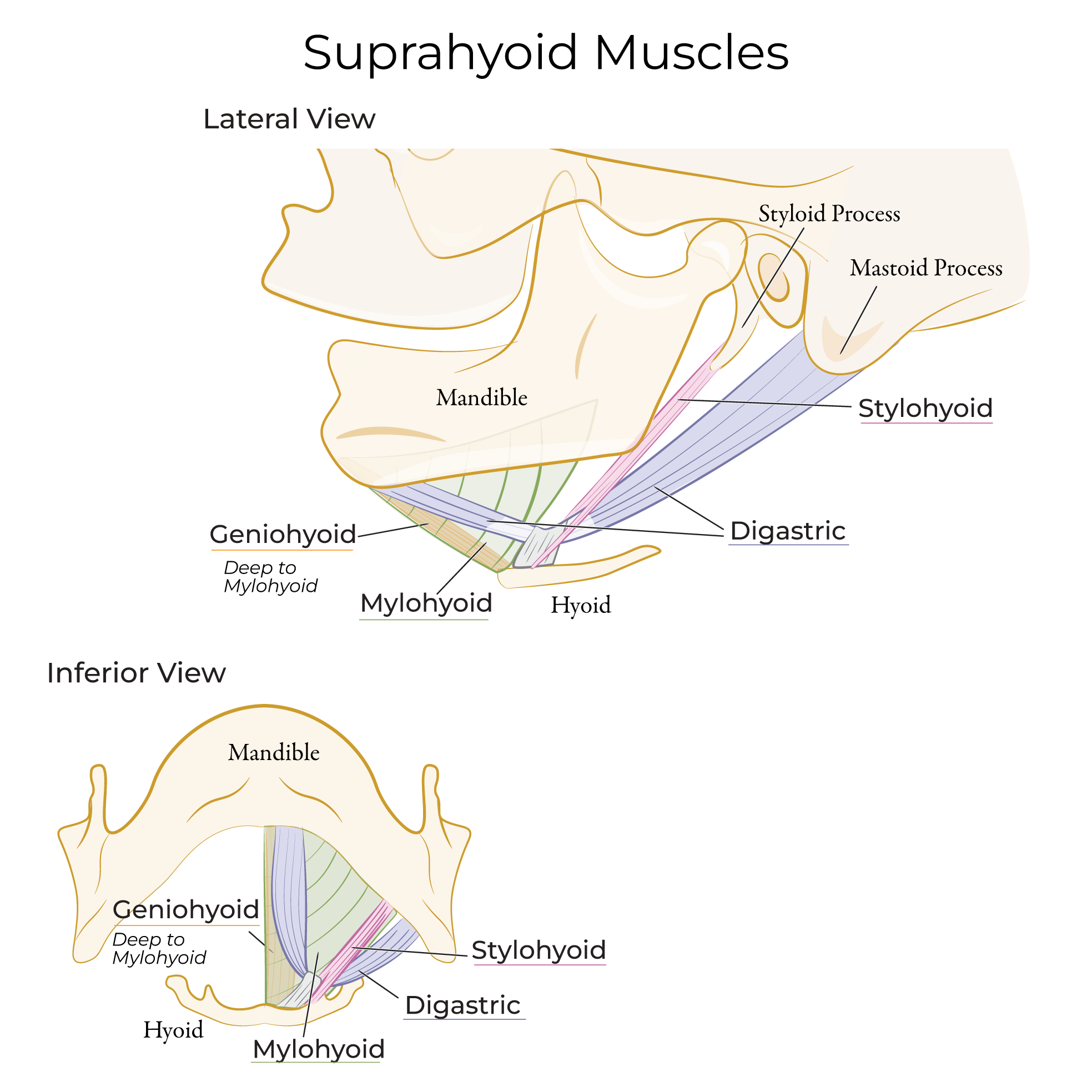
Show additional boundaries of the oral cavity: the mandible and lower teeth and the muscles of the floor of the mouth, the tongue, geniohyoid, and mylohyoid.
Now, show the thyroid cartilage, epiglottis, and the internal surface of the larynx; highlight the laryngeal inlet. Show the connective tissues between the hyoid bone and the thyroid cartilage and epiglottis.
Indicate the pharyngeal constrictors, which form the half-cylinder muscular tube of the pharynx: label them as superior, middle, and inferior pharyngeal constrictors.
Show that the internal surface of the constrictors is covered and supported by the pharyngobasilar fascia; the external surface of the constrictors is covered by the thinner buccopharyngeal fascia.
Three regions of the pharynx:
The nasopharynx opens to the nasal cavity.
The oropharynx opens to the oral cavity,
And, the laryngopharynx opens to the larynx.
Mucosal Features
Palatoglossal fold which covers the palatoglossus muscle, and the palatine tonsil. The palatoglossus muscle elevates the tongue and pulls the soft palate inferiorly, narrowing the oropharyngeal isthmus. Located in the oropharynx.
Pharyngeal tonsil in the roof of the nasopharynx.
Pharyngotympanic tube opening near the pharyngeal tonsil. The opening produces an elevation called the torus tubularis.
Torus levatorius is the mucosal fold over levator veli palatini.
Salpingopharyngeal fold is the mucosa over the salpingopharyngeus.
Palatopharyngeal arch is the mucosal fold that overlies the palatopharyngeal muscle.
Posterior View
Set up the diagram.
First, draw the base of the skull, the hyoid bone, larynx, and trachea.
Now draw the splayed-open mucosa.
Mucosal Features
Openings to anterior structures:
The choanae is where the nasopharynx opens to the nasal cavity
The oropharyngeal isthmus is where the oropharynx opens to the oral cavity – show that we can see the pharyngeal portion of the tongue,
The laryngeal inlet the laryngopharynx opens to the larynx.
Show that the pharynx is continuous with the esophagus.
In the nasopharynx, indicate the following:
The pharyngeal recess, which is a deep depression posterior to the opening of the pharyngotympanic tube.
Pharyngeal tonsil at the roof of the nasopharynx.
On the sides of the choanae, show the torus tubularis (again, this is the elevation where the pharyngotympanic tube opens to the pharynx); show that the salpingopharyngeal fold extends vertically from the tubularis.
Torus levatorius (the mucosal fold over levator veli palatini).
Area of the soft palate (comprises the palatine aponeurosis and muscles).
In the oropharynx, indicate the palatopharyngeal arch (the mucosal fold that overlies the palatopharyngeal muscle).
Show the palatine tonsil and, in the pharyngeal portion of the tongue, show the lingual tonsil.
Lastly, in the laryngopharynx, show the piriform fossa (aka sinus; also spelled pyrifirom) lies to the side of the larynx; this is a common site of hypopharyngeal cancer.