

Start your One-Week Free Trial
Already subscribed? Log in »


Oral Cavity
Key Definitions
Anatomical boundaries:
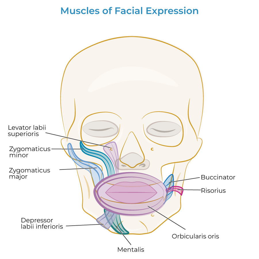
Anteriorly, the oral cavity is bound by the lips (orbicularis oris and other muscles that insert on the lips).
Posteriorly, the oral cavity is open to the oropharynx.
Laterally, the cavity is bound by the cheeks.
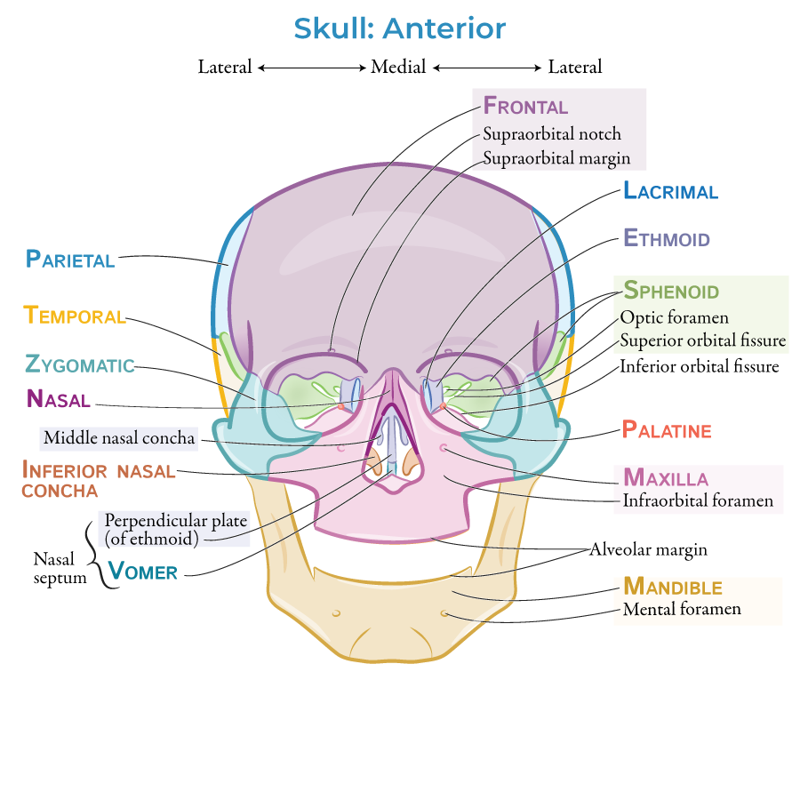
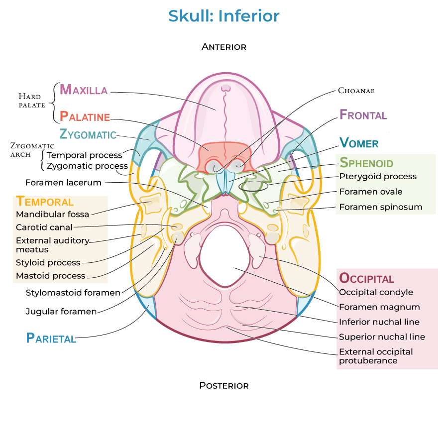
Superiorly, the "roof" of the oral cavity is formed by the hard palate (the maxillary and palatine bones) and the soft palate (the aponeurosis and muscles); we call this the "roof" of the mouth.
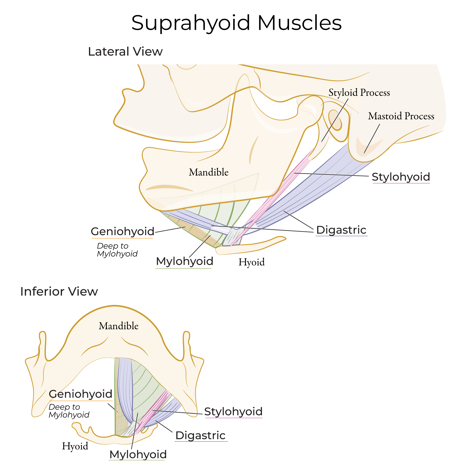
Inferiorly, the mylohyoid and geniohyoid muscles form the "floor" of the mouth.
Digestive Functions:
The teeth and tongue physically manipulate foods, and salivary enzymes chemically digest carbohydrates. The oral cavity is also involved in speech, particularly in sound modification.
Anterior View
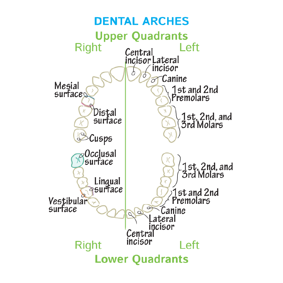
 The gingivae (the gums) and the teeth; the upper teeth are housed in the maxilla bone and the lower teeth are housed in the mandible (aka jaw bone).
 The superior and inferior labial frenula are folds of the mucous membrane; if you push your tongue between your lips and front teeth, you can feel this.
The tongue and the lingual frenulum, which is a mucosal fold that attaches the inferior surface of the tongue to the floor of the oral cavity.
Ankyloglossia (aka tongue-tie) is a developmental anomaly in which the lingual frenulum is unusually short or thick, limiting the tongue's range of motion. In infancy, this can make feeding (especially breastfeeding) difficult and lead to undernourishment.
Salivary duct orifices open under the tongue.
The hard palate comprises most of the roof of the mouth, and the soft palate makes up the posterior portion.
The hard palate is formed by the maxillary and palatine bones.
The superior and inferior labial frenula are folds of the mucous membrane; if you push your tongue between your lips and front teeth, you can feel this.
The tongue and the lingual frenulum, which is a mucosal fold that attaches the inferior surface of the tongue to the floor of the oral cavity.
Ankyloglossia (aka tongue-tie) is a developmental anomaly in which the lingual frenulum is unusually short or thick, limiting the tongue's range of motion. In infancy, this can make feeding (especially breastfeeding) difficult and lead to undernourishment.
Salivary duct orifices open under the tongue.
The hard palate comprises most of the roof of the mouth, and the soft palate makes up the posterior portion.
The hard palate is formed by the maxillary and palatine bones.
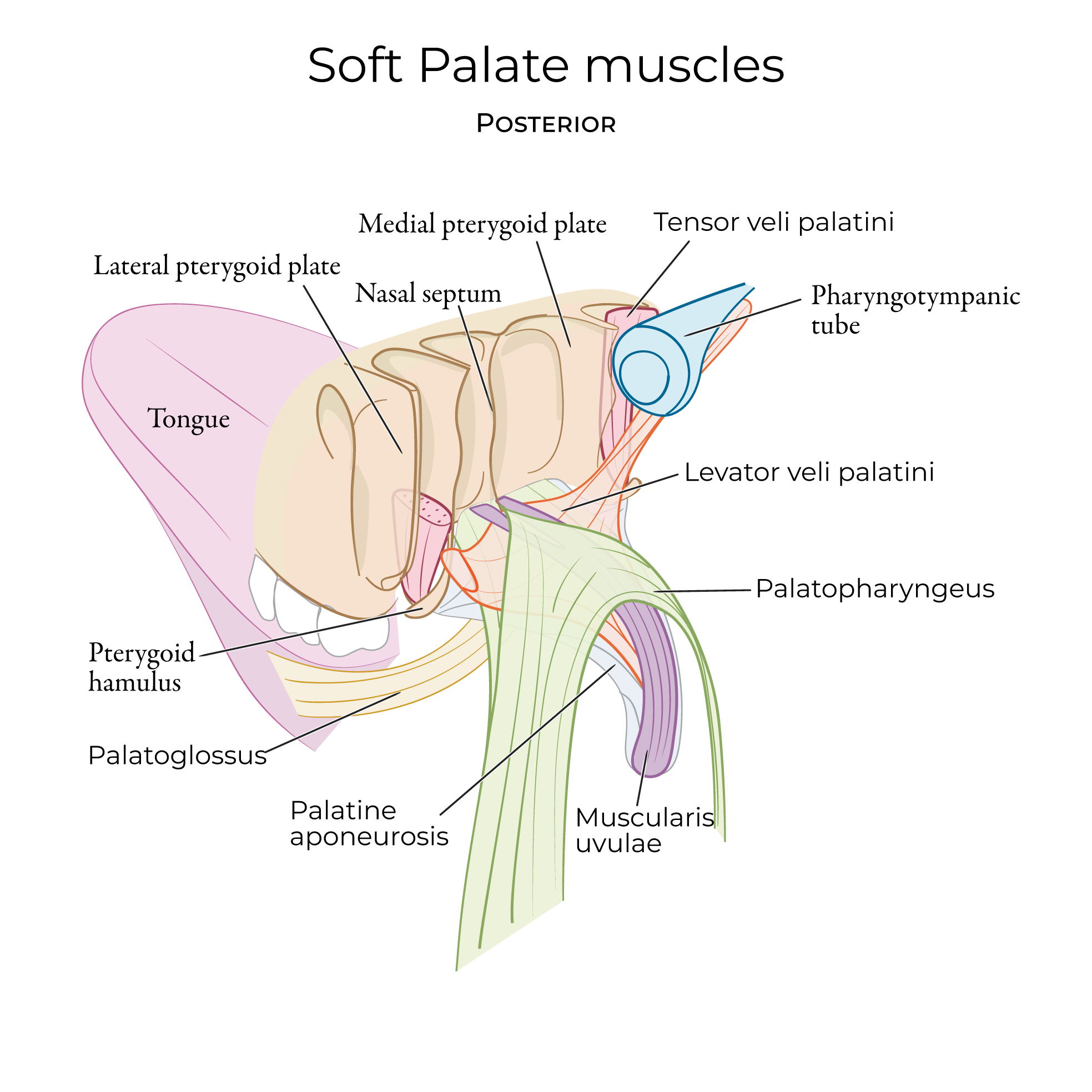
 The soft palate is formed by the palatine aponeurosis and muscles.
 The uvula is the posterior free edge of the soft palate; during swallowing, the soft palate elevates and prevents foods and liquids from entering the nasal cavity.
The midline palatine raphe indicates where the right and left sides of the palate fused during fetal development; you can feel this ridge if you run your tongue along the roof of your mouth.
Failure of the right and left sides of the palate to fuse results in a cleft palate, which is characterized by an opening in the palate at birth that interferes with eating and speaking.
On either side of the palatine raphe, we show the transverse palatine folds (aka palatine rugae), which are ridges of connective tissue that create friction and aid in bolus formation during the oral phase of digestion.
The palatoglossal arch extends from the soft palate to the tongue; it contains the palatoglossal muscle.
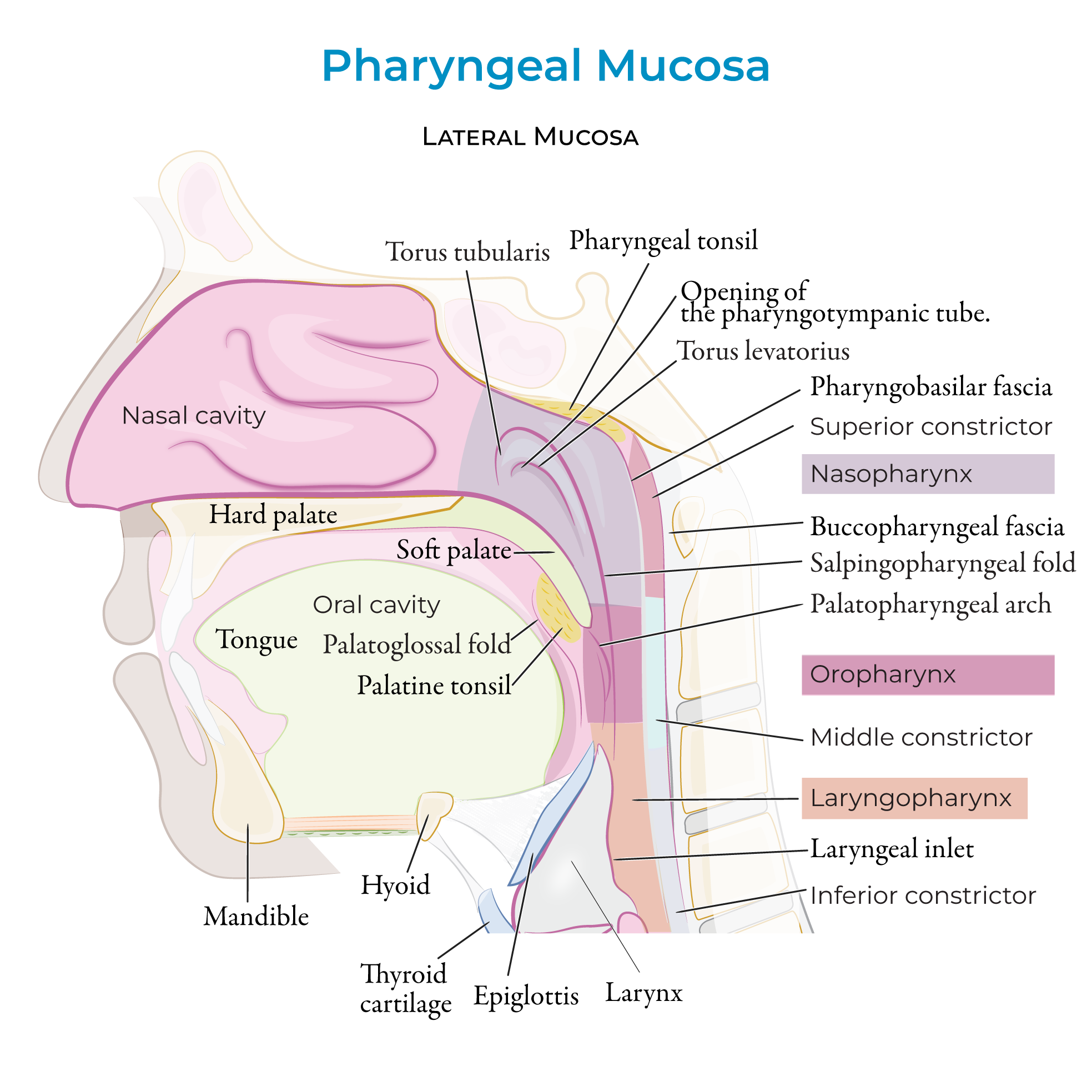
Posterior to this, show the palatopharyngeal arch, which contains the palatopharyngeal muscle.
Between the two arches, show the palatine tonsils, which are collections of lymphoid tissue that monitor and respond to ingested microbes.
The fauces is the posterior opening of the oral cavity; foods and liquids exit the oral cavity via the fauces and enter the pharynx.
The uvula is the posterior free edge of the soft palate; during swallowing, the soft palate elevates and prevents foods and liquids from entering the nasal cavity.
The midline palatine raphe indicates where the right and left sides of the palate fused during fetal development; you can feel this ridge if you run your tongue along the roof of your mouth.
Failure of the right and left sides of the palate to fuse results in a cleft palate, which is characterized by an opening in the palate at birth that interferes with eating and speaking.
On either side of the palatine raphe, we show the transverse palatine folds (aka palatine rugae), which are ridges of connective tissue that create friction and aid in bolus formation during the oral phase of digestion.
The palatoglossal arch extends from the soft palate to the tongue; it contains the palatoglossal muscle.
Posterior to this, show the palatopharyngeal arch, which contains the palatopharyngeal muscle.
Between the two arches, show the palatine tonsils, which are collections of lymphoid tissue that monitor and respond to ingested microbes.
The fauces is the posterior opening of the oral cavity; foods and liquids exit the oral cavity via the fauces and enter the pharynx.
Sagittal View
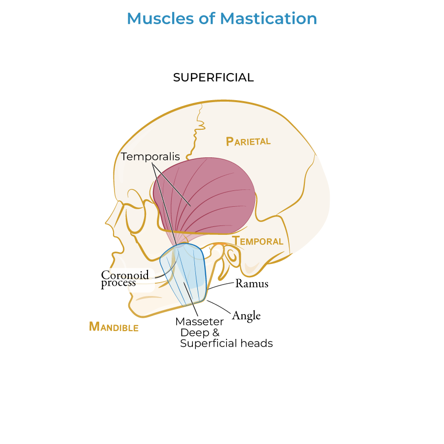
 Then show the posterior portion of buccinator, the flat sheet-like muscle of the cheek.
Masseter runs vertically between the mandible and zygomatic bones.
Then show the posterior portion of buccinator, the flat sheet-like muscle of the cheek.
Masseter runs vertically between the mandible and zygomatic bones.
 The tongue fills the closed oral cavity and is connected to the oral mucosa via the lingual frenulum; posteriorly, its root is attached to the hyoid bone. The tongue comprises 4 intrinsic and 4 extrinsic muscles that change its shape and positioning for speech and eating/drinking.
Minor salivary glands are located throughout the oral submucosa.
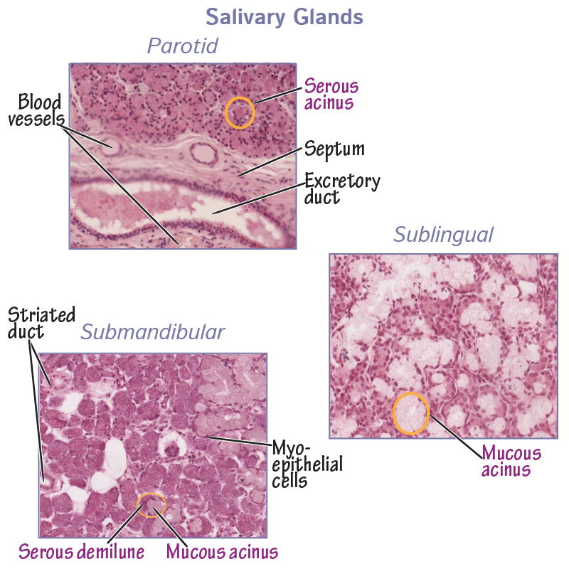
Indicate the three major salivary glands as follows:
The parotid gland is the largest, it resides in the side of face just anterior to the ear and superficial to the masseter muscle. The parotid gland has a long duct that drains saliva from the parotid gland to the mouth. This duct is large and easily seen during dissection.
The sublingual gland lies beneath the tongue in the floor of the mouth. It secretes saliva via several small ducts.
The submandibular gland lies along the medial side of the mandible and wraps around the mylohyoid muscle; its duct also drains into the mouth.
The tongue fills the closed oral cavity and is connected to the oral mucosa via the lingual frenulum; posteriorly, its root is attached to the hyoid bone. The tongue comprises 4 intrinsic and 4 extrinsic muscles that change its shape and positioning for speech and eating/drinking.
Minor salivary glands are located throughout the oral submucosa.
Indicate the three major salivary glands as follows:
The parotid gland is the largest, it resides in the side of face just anterior to the ear and superficial to the masseter muscle. The parotid gland has a long duct that drains saliva from the parotid gland to the mouth. This duct is large and easily seen during dissection.
The sublingual gland lies beneath the tongue in the floor of the mouth. It secretes saliva via several small ducts.
The submandibular gland lies along the medial side of the mandible and wraps around the mylohyoid muscle; its duct also drains into the mouth.
 The salivary glands are important for oral cavity lubrication.
If salivary production is decreased, patients experience difficulty eating and speaking, and their rate of dental decay increases.
The salivary glands are important for oral cavity lubrication.
If salivary production is decreased, patients experience difficulty eating and speaking, and their rate of dental decay increases.
Tongue Muscles
Intrinsic Muscles
Originate and insert within the tongue itself.
The superior longitudinal muscle, the vertical and transverse muscle, and the inferior longitudinal muscle.
These muscles lengthen and shorten the tongue, curl it along its apex and edges, and flatten and round its surface.
Extrinsic muscles
Protrude and retract, depress and elevate the tongue.
The first three muscles insert laterally on the tongue:
Palatoglossus, which is a muscle of the soft palate.
Styloglossus, which arises from the styloid process of the skull.
Hyoglossus, which arises from the hyoid bone.
Genioglossus is a thick fan-shaped muscle that inserts along the dorsum of the intrinsic tongue muscles; its bony anchors are the mandible and hyoid bone.
Genioglossus contributes significantly to the body of the tongue.
Innervation
Seven of the 8 tongue muscles are innervated by the hypoglossal nerve (CN XII); palatoglossus, the outlier, is innervated by the vagus nerve (CN X).
Thus, asking a patient to stick their tongue out tests CN XII functioning: if there is a nerve lesion, the tip of the tongue will deviate towards the lesion.