

Start your One-Week Free Trial
Already subscribed? Log in »

Anterior Thigh Muscles
Here we'll learn the muscles of the anterior thigh, which flex the hip joint and extend the knee.
Femur
Pelvic Girdle
Origins & Insertions
Arteries
Innervation
Photos


 Comprises rectus femoris, vastus medialis, vastus lateralis, and vastus intermedius.
– These muscles insert at the patella and tibial tuberosity (via common tendon of quadriceps femoris).
– They all extend the leg.
Rectus femoris has two origins:
– Originates on the anterior inferior iliac spine and the superior rim of the acetabulum.
– Rectus femoris flexes the thigh (in addition to extending the leg like the other quadriceps muscles).
– Note that it crosses both the hip and the knee, which is why it acts on both the femur and the leg.
Vastus medialis
– Originates on the intertrochanteric line and linea aspera (of the femur).
Vastus lateralis
– Originates on the intertrochanteric line, linea aspera, greater trochanter, and gluteal tuberosity.
Vastus intermedius
– Originates on the shaft of the proximal femur.
Comprises rectus femoris, vastus medialis, vastus lateralis, and vastus intermedius.
– These muscles insert at the patella and tibial tuberosity (via common tendon of quadriceps femoris).
– They all extend the leg.
Rectus femoris has two origins:
– Originates on the anterior inferior iliac spine and the superior rim of the acetabulum.
– Rectus femoris flexes the thigh (in addition to extending the leg like the other quadriceps muscles).
– Note that it crosses both the hip and the knee, which is why it acts on both the femur and the leg.
Vastus medialis
– Originates on the intertrochanteric line and linea aspera (of the femur).
Vastus lateralis
– Originates on the intertrochanteric line, linea aspera, greater trochanter, and gluteal tuberosity.
Vastus intermedius
– Originates on the shaft of the proximal femur.
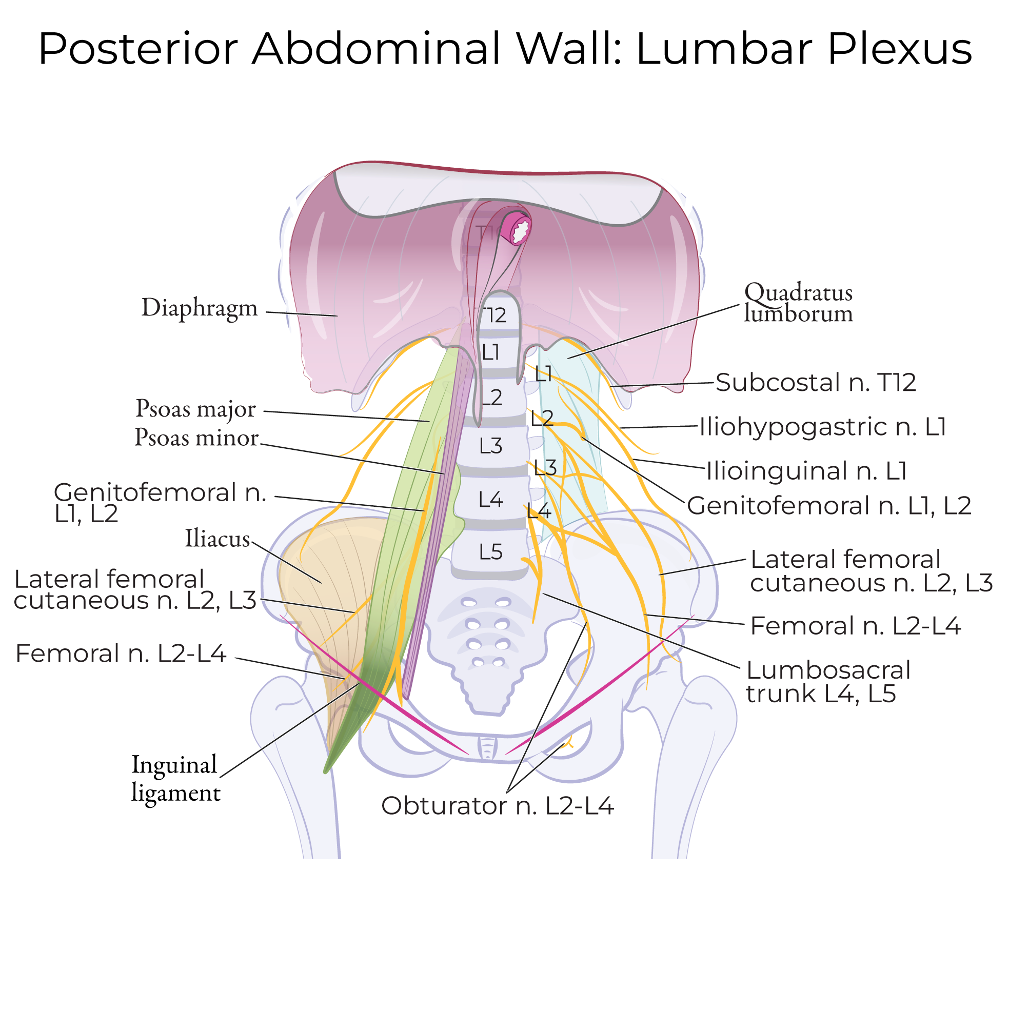
 Innervation in primarily via the femoral nerve, which innervates quadriceps femoris, sartorius, and iliacus.
The anterior rami of lumbar nerves L1-L3 innervate psoas major.
Innervation in primarily via the femoral nerve, which innervates quadriceps femoris, sartorius, and iliacus.
The anterior rami of lumbar nerves L1-L3 innervate psoas major.
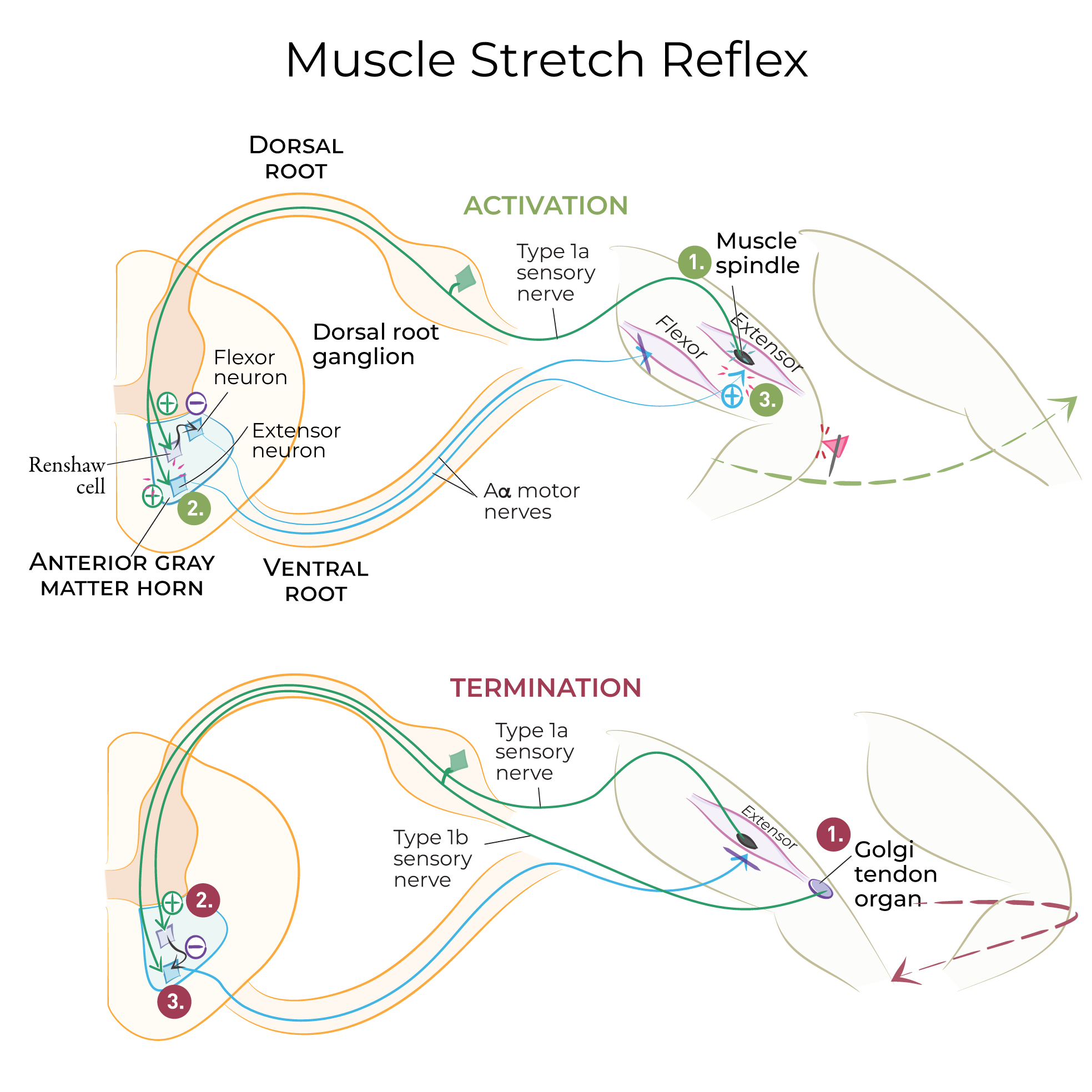
 The patellar reflex test is used to assess the L2, L3, and L4 segments of the spinal cord. In response to tapping the patellar tendon, a rapid, involuntary contraction of the quadriceps muscles kicks the leg forward.
Review & Additional Images
Muscles


Iliospoas
Comprises iliacus and psoas major:
- Iliacus arises from the iliac fossa.
- Psoas major arises from vertebrae T12-L5.
- Together, these muscles insert on the lesser trochanter, and flex the thigh.

- Originates on the anterior superior iliac spine
- Inserts on the proximal anteromedial tibia.
- It flexes and laterally rotates the thigh; flexes and medially rotates the leg.
- Most superficial muscle of the anterior thigh; crosses over quadriceps femoris.
 Comprises rectus femoris, vastus medialis, vastus lateralis, and vastus intermedius.
– These muscles insert at the patella and tibial tuberosity (via common tendon of quadriceps femoris).
– They all extend the leg.
Rectus femoris has two origins:
– Originates on the anterior inferior iliac spine and the superior rim of the acetabulum.
– Rectus femoris flexes the thigh (in addition to extending the leg like the other quadriceps muscles).
– Note that it crosses both the hip and the knee, which is why it acts on both the femur and the leg.
Vastus medialis
– Originates on the intertrochanteric line and linea aspera (of the femur).
Vastus lateralis
– Originates on the intertrochanteric line, linea aspera, greater trochanter, and gluteal tuberosity.
Vastus intermedius
– Originates on the shaft of the proximal femur.
Comprises rectus femoris, vastus medialis, vastus lateralis, and vastus intermedius.
– These muscles insert at the patella and tibial tuberosity (via common tendon of quadriceps femoris).
– They all extend the leg.
Rectus femoris has two origins:
– Originates on the anterior inferior iliac spine and the superior rim of the acetabulum.
– Rectus femoris flexes the thigh (in addition to extending the leg like the other quadriceps muscles).
– Note that it crosses both the hip and the knee, which is why it acts on both the femur and the leg.
Vastus medialis
– Originates on the intertrochanteric line and linea aspera (of the femur).
Vastus lateralis
– Originates on the intertrochanteric line, linea aspera, greater trochanter, and gluteal tuberosity.
Vastus intermedius
– Originates on the shaft of the proximal femur.

Anterior thigh innervation
Patellar reflex test
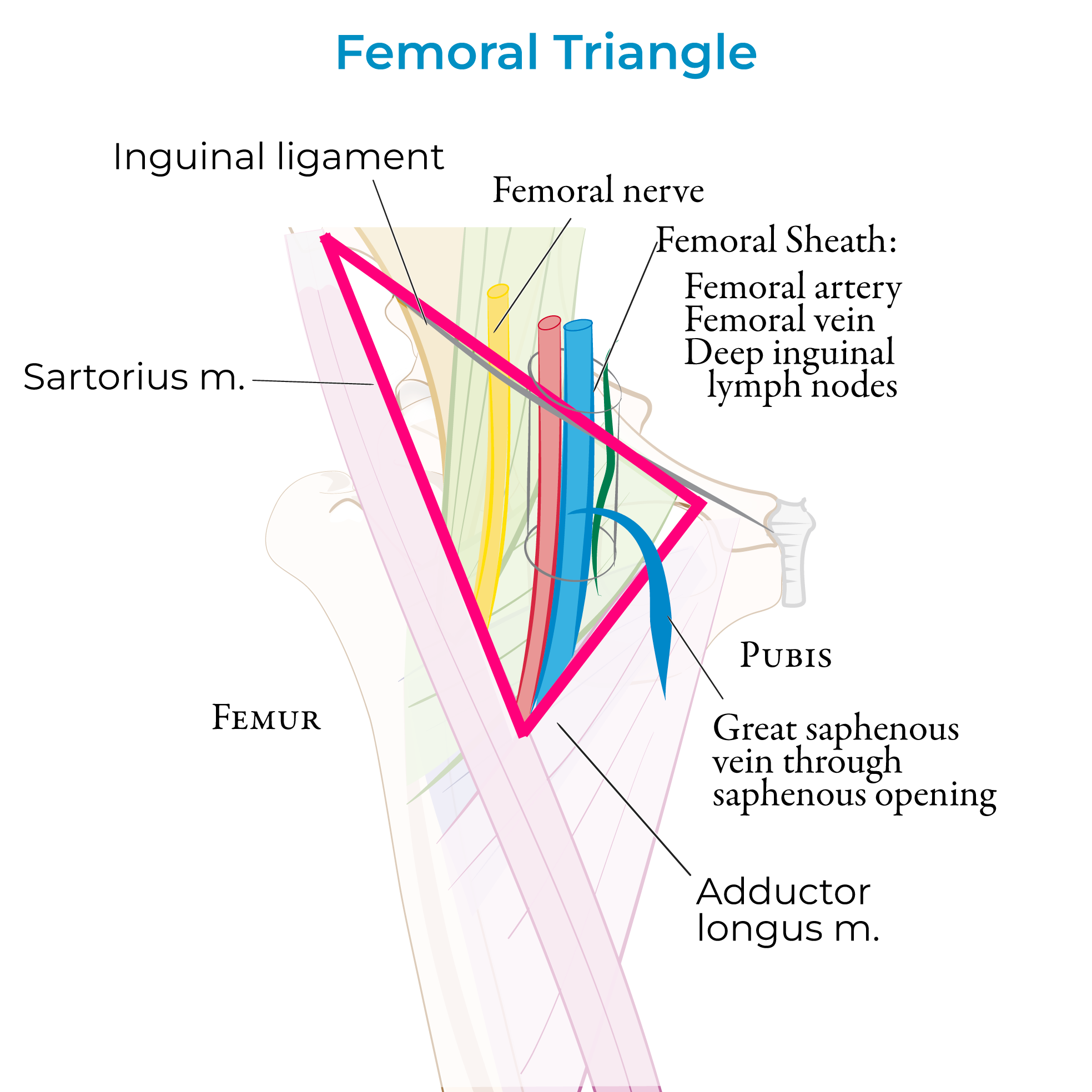
Femoral Triangle
Femoral Triangle Boundaries
The inguinal ligament forms the superior boundary; it forms the base of the triangle.
The lateral border of adductor longus forms the medial border.
Sartorius forms the lateral border of the triangle; the apex is formed by the crossing of sartorius over adductor longus.
The floor of the triangle comprises the muscles iliopsoas and pectineus.
The roof of the triangle is formed by the fascia lata of the thigh (along with the cribriform fascia, subcutaneous tissues, and skin of the thigh). The fascia lata comprises a thick band of connective tissue that envelops the muscles of the thigh.
Contents of the Femoral Triangle
Neurovascular and lymphatic structures pass through the space deep to the inguinal ligament between the pelvis and the thigh; this space is called the retro-inguinal space.
The vessels bisect the femoral triangle on their way to the adductor canal to traverse the lower thigh.
Mnemonic, from lateral to medial
N - Femoral Nerve and terminal branches.
A - Femoral Artery and its branches.
V - Femoral Vein
E - Empty space (the femoral canal – see notes below)
L - Deep Lymph nodes
Femoral Sheath:
The femoral sheath is a 3-4 cm long fascial tube derived from transversalis and iliac fascia. The sheath envelops the femoral artery and vein and their branches, as well as the deep inguinal lymph nodes.
The fascial sheath itself is divided into compartments:
Lateral, containing the femoral artery.
Intermediate, containing the femoral vein.
Medial, which houses the femoral canal with lymph vessels.
Great Saphenous Vein & Clinical Correlations
The great saphenous vein, which is the long superficial vein that drains the lower extremity, passes through the saphenous opening of the fascia lata to drain into the femoral vein within the femoral triangle.
The great saphenous vein is often used in arterial bypass operations (coronary artery and peripheral), due to its large size and relatively easy access.
In an emergency setting, the great saphenous vein can be used for venous cutdown, in which the vein is exposed and a cannula or catheter is inserted into the vessel.
Femoral Hernias
At the proximal end of the femoral canal, we'll find the femoral ring - this is a clinically relevant area because femoral hernias occur when abdominal tissues, such as the intestines, protrude through the femoral ring to enter the femoral canal.
Femoral hernias are caused by straining that increases intra-abdominal pressure, as when coughing, heavy lifting, defecating or urinating, pregnancy and childbirth, etc.
Femoral hernias are rare overall but require surgical repair due to their high rate of bowel obstruction and ischemia.