

Start your One-Week Free Trial
Already subscribed? Log in »

Testes and Supportive Tract Development
Here we will learn the embryology of the testes and the associated ducts and glands.
We will start our diagram in approximately week 5, when the primordial germ cells migrate to the gonadal ridges.
To show this, we draw the lower portion of the embryo, with the aorta, dorsal mesentery, hindgut, yolk stalk, and mesonephros; indicate the gonadal ridges along the posterior body wall, medial to the mesonephros.
The primordial germ cells exit the yolk stalk, migrate through the dorsal mesentery, and arrive at the gonadal ridges.
In an XY gonad, the SRY gene, which is located on the Y chromosome, initiates the transformation of the indifferent gonad into a testis.
Supporting-cell precursors in the testis express the SRY gene, which activates SOX9 and drives Sertoli-cell differentiation in the nearby cells (note that Sertoli cells are also called sustentacular cells).
The Sertoli cells “recruit” other cells that transform to become Leydig (aka, interstitial) cells; the Leydig cells produce testosterone, which plays a role in male reproductive tract formation during gestation. Eventually, these fetal Leydig cells will disappear, and a new adult generation will form during puberty, when full maturation of the reproductive system occurs.
Later in development, we'll see interstitial Leydig cells and testis cords; the primordial germ cells and Sertoli cells are reorganized to form the cords.
Let’s “cut” one of these cords to show it in cross-section: indicate the primordial germ cells in the center, surrounded by Sertoli cells; the outermost layer of the cord comprises peritubular myoid cells.
Later in life, the peritubular myoid cells contract to propel the contents of the cords.
The testis cords remain solid until puberty, when they are canalized to form the seminiferous tubules; likewise, the primordial germ cells are developmentally arrested until onset of meiosis during puberty.
The formation of the reproductive tract and accessory gland formation occurs during weeks 8-12.
First, we show how the gonads are situated in relation to the mesonephric and paramesonephric ducts.
Indicate the location of the urogenital sinus; recall it is derived from endoderm; superiorly, we show the location of the gonads (the early testes).
The paramesonephric, aka, Mullerian ducts emerge lateral to the gonads, cross over the mesonephric ducts, and run caudally towards the urogenital sinus.
Anti-Mullerian hormone (AMH) causes regression of the paramesonephric ducts; AMH is produced by the Sertoli cells of the testis (small, non-functional remnants of the paramesonephric ducts may appear as the appendix testis).
Testosterone produced by the Leydig cells of the testes maintains the mesonephric structures, which are incorporated into the developing reproductive tract.
First, we draw a mesonephric duct and its tubules; the testis comprises the solid testis cords and interstitial cells.
Show that testosterone induces the following changes:
The outer layer thickens and is now called the tunica albuginea; it covers the solid testis cords. The mesonephric tubules give rise to efferent ductules; the mesonephric duct gives rise to the epididymis, which is a tightly coiled tube and to the ductus deferens (aka, vas deferens). The rete testis has dual origins, arising from the mesonephric tubules and the Sertoli cells.
After canalization of the testis cords, sperm cells and seminal fluids will pass from the seminiferous tubules, through the rete testis and efferent ductules, into the epididymis and to the ductus deferens, which will ultimately deliver the seminal products to the urethra for ejaculation.
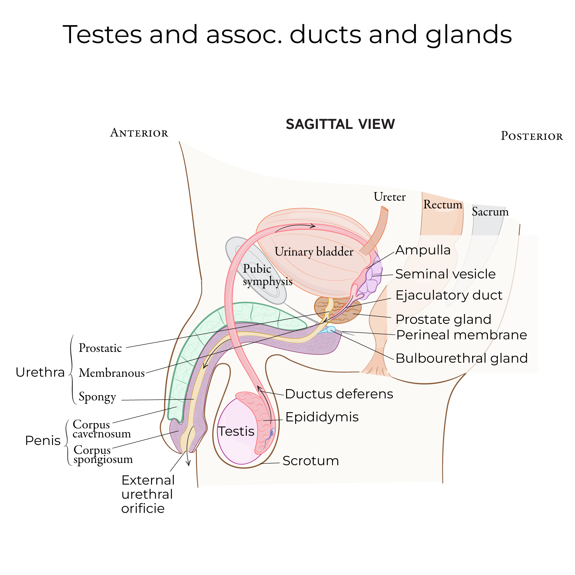
Finally, we show the connection of these ducts and tubules to the urethra and the formation of the male accessory glands.
First, we indicate the urinary bladder and urethra; posteriorly, we see the distal end of the ductus deferens, which empties into the urethra via the ejaculatory duct.
The seminal vesicles arise as outgrowths of the distal mesonephric duct near the future ejaculatory duct. Notice that the contents of both the ductus deferens and the seminal vesicles will pass through the ejaculatory duct to reach the urethra.
Prostatic tissue arises from the endoderm of the urethra as a series of outgrowths that eventually form the singular gland. We label the prostatic portion of the urethra, which passes through the prostate and receives its secretions, and the membranous portion, which passes through the pelvic diaphragm. The penile urethra passes through the penis.
 The primordial germ cells give rise to the spermatogonia, the early-stage testicular gametes.
The genital ridge and coelomic epithelium give rise to the Leydig and Sertoli cells.
The mesonephros gives rise to the rete testis (which also has Sertoli cell origins), ductus deferens, epididymis, and seminal vesicles (these last three specifically arise from the mesonephric duct).
And, the endoderm of the urogenital sinus gives rise to the prostate and bulbourethral glands (not shown in our diagram).
The primordial germ cells give rise to the spermatogonia, the early-stage testicular gametes.
The genital ridge and coelomic epithelium give rise to the Leydig and Sertoli cells.
The mesonephros gives rise to the rete testis (which also has Sertoli cell origins), ductus deferens, epididymis, and seminal vesicles (these last three specifically arise from the mesonephric duct).
And, the endoderm of the urogenital sinus gives rise to the prostate and bulbourethral glands (not shown in our diagram).
Primordial Germ Cells
Associated Tracts and Glands
Summary