

Start your One-Week Free Trial
Already subscribed? Log in »

Testes Anatomy & Histology
Testes
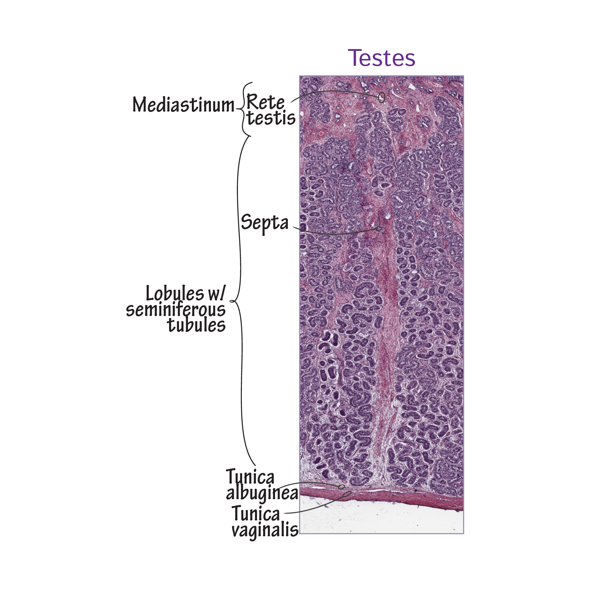 Immature sperm exit the seminiferous tubules via straight tubules, which deliver them to the rete testis in the mediastinum.
The rete testis concentrates non-motile sperm before they are moved to the epididymis via the efferent ductules.
The epididymis comprises a single tightly coiled duct; sperm are moved through the head, body, and tail, with fluid reabsorption and concentration continuing throughout. Sperm cells are stored in the epididymis for approximately two weeks, during which time they achieve motility.
The tail of the epididymis is continuous with the ductus deferens, which transports the sperm through the spermatic cord and ultimately to the ejaculatory duct, where the sperm cells join with various fluids to form semen.
The tunica vaginalis is a double-layered sac that comprises a visceral layer that overlies the tunica albuginea, and a parietal layer, which is in contact with the innermost layer of the scrotum. Label the cavity between the visceral and parietal layers; this potential space contains a small amount of fluid, which allows for movement of the testicles within the scrotum.
Immature sperm exit the seminiferous tubules via straight tubules, which deliver them to the rete testis in the mediastinum.
The rete testis concentrates non-motile sperm before they are moved to the epididymis via the efferent ductules.
The epididymis comprises a single tightly coiled duct; sperm are moved through the head, body, and tail, with fluid reabsorption and concentration continuing throughout. Sperm cells are stored in the epididymis for approximately two weeks, during which time they achieve motility.
The tail of the epididymis is continuous with the ductus deferens, which transports the sperm through the spermatic cord and ultimately to the ejaculatory duct, where the sperm cells join with various fluids to form semen.
The tunica vaginalis is a double-layered sac that comprises a visceral layer that overlies the tunica albuginea, and a parietal layer, which is in contact with the innermost layer of the scrotum. Label the cavity between the visceral and parietal layers; this potential space contains a small amount of fluid, which allows for movement of the testicles within the scrotum.