
PANCE - H. pylori Infection
Start your One-Week Free Trial
Already subscribed? Log in »
Here are key facts for PANCE from the Helicobacter pylori tutorial, as well as points of interest at the end of this document that are not directly addressed in this tutorial but should help you prepare for the boards.
 Below is information not explicitly contained within the tutorial but important for PANCE.
- --
VITAL FOR PANCE
Helicobacter pylori - Organism Characteristics
1. H. pylori is a spiral, Gram-negative rod that can appear as coccoid in older cultures.
2. It is catalase, oxidase, and urease positive - critical for diagnostic testing.
3. Microaerobic organism that grows in conditions of reduced oxygen and increased carbon dioxide.
4. Transmission is human-to-human, with infection typically occurring during childhood.
5. Creates life-long colonization with symptoms often presenting during adulthood.
Clinical Presentation
1. Gastritis presents with inflammation of the stomach lining and infiltration of neutrophils and mononuclear cells.
2. Patients may be asymptomatic or experience an acute phase of nausea, bloating, and vomiting.
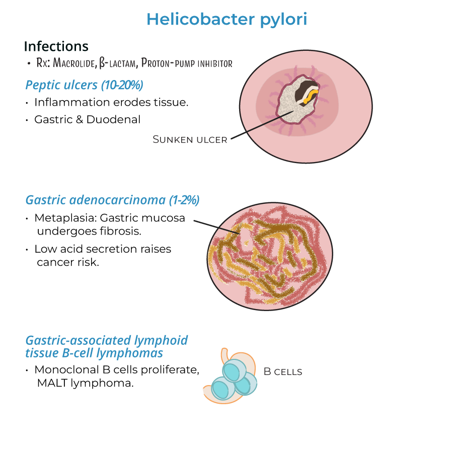
3. Peptic ulcers develop in 10-20% of gastritis patients, occurring in either stomach or duodenum.
4. Gastric adenocarcinoma develops in approximately 1-2% of chronic infections.
5. MALT lymphomas form when monoclonal B cells proliferate in gastric lymphoid tissue.
Diagnostic Considerations
1. Urease activity is a key diagnostic marker for H. pylori infection.
2. Inflammation can be localized (usually to pyloric antrum) or widespread (pangastritis).
3. Antral gastritis associates with increased acid production and duodenal ulcers.
4. Pangastritis (multifocal inflammation) associates with atrophy, reduced acid production, and gastric cancer risk.
Treatment Approach
1. Standard therapy includes macrolides, beta-lactams, and proton-pump inhibitors.
2. Treatment is essential because chronic gastritis can lead to severe complications.
- --
HIGH YIELD
Virulence Factors and Pathogenesis
1. Urease converts urea to ammonia and bicarbonate to neutralize gastric acids.
2. Multiple flagella provide corkscrew motility to penetrate gastric mucus.
3. Mucinase production allows bacteria to migrate through viscous stomach mucus.
4. Infection triggers host production of IL-8, recruiting neutrophils that damage host tissues.
5. Bacteria protect themselves by producing superoxide dismutase and catalase.
6. Vacuolating cytotoxin A (VacA) promotes pore formation and induces host cell death.
7. Cytotoxin-associated gene A (CagA) promotes tissue changes and T-cell apoptosis.
8. Type IV secretion systems inject CagA effector protein into host cells.
Disease Progression
1. H. pylori destroys mucosa, allowing acids and toxins access to deeper tissues.
2. Chronic inflammation leads to metaplasia where gastric mucosa is replaced by fibrotic tissue.
3. Reduced gastric acid secretion is associated with higher risk of adenocarcinoma.
4. Severe ulceration can lead to bleeding, perforation, and metaplasia.
5. In MALT lymphoma, lymphoid tissues infiltrate the stomach with monoclonal B-cell proliferation.
Clinical Variants
1. Ulcers can occur in either the stomach or duodenum (first portion of small intestine).
2. Gastritis can be localized or widespread, affecting treatment approach and prognosis.
3. T-helper 1 cells are implicated in the inflammatory response to H. pylori.
Special Considerations
1. Enterohepatic helicobacters (H. cinaedi and H. fennelliae) cause gastroenteritis and bacteremia.
2. These related species primarily affect immunocompromised individuals.
3. Unlike H. pylori, these species invade the intestines and liver rather than the stomach.
- --
Beyond the Tutorial
Clinical Assessment and Diagnosis
1. Indications for testing: persistent dyspepsia, peptic ulcer disease, gastric MALT lymphoma, history of gastric cancer.
2. Non-invasive testing options: urea breath test, stool antigen test, serologic testing.
3. Invasive testing: endoscopy with biopsy for rapid urease test, histology, and culture.
4. Alarm features requiring immediate endoscopy: age >55 with new-onset symptoms, weight loss, dysphagia, persistent vomiting, GI bleeding, iron deficiency anemia, palpable mass.
5. Differential diagnosis: functional dyspepsia, GERD, gastric cancer, medication-induced gastritis, autoimmune gastritis.
Treatment Guidelines
1. First-line therapy: clarithromycin triple therapy (clarithromycin, amoxicillin/metronidazole, PPI) in low-resistance areas.
2. Alternative first-line: bismuth quadruple therapy (bismuth, tetracycline, metronidazole, PPI).
3. Second-line options: levofloxacin triple therapy, rifabutin-based therapy.
4. Treatment duration: 14 days preferred over 10 or 7 days.
5. Post-treatment testing: urea breath test or stool antigen test at least 4 weeks after completion of therapy.
6. Treatment of refractory cases: culture with sensitivity testing, alternative antibiotic combinations.
Prevention and Patient Education
1. Lifestyle modifications: smoking cessation, alcohol reduction, avoidance of NSAIDs.
2. Family screening in patients with gastric cancer history.
3. Testing before long-term PPI therapy.
4. Counseling on medication adherence to ensure eradication.
5. Follow-up recommendations based on findings and risk stratification.
Emerging Concepts
1. Antibiotic resistance patterns and impact on treatment selection.
2. Role of probiotics as adjunctive therapy.
3. Potential protective effects against esophageal diseases (controversial).
4. Association with extraintestinal manifestations: ITP, iron deficiency anemia, vitamin B12 deficiency.
5. Relationship between H. pylori and the gut microbiome.