
ABIM - Myocardial Infarction Symptoms, Diagnosis, & Treatments
Start your One-Week Free Trial
Already subscribed? Log in »
Here are key facts for American Board of Internal Medicine (ABIM) Examination from the Myocardial Infarctions: Diagnosis & Treatment tutorial, focusing on clinical management and treatment decision-making that are essential for board certification. See the tutorial notes for further details and relevant links.
 Below is information not explicitly contained within the tutorial but important for the American Board of Internal Medicine Examination.
- --
VITAL FOR ABIM
Epidemiology & Disparities in Care
1. Global patterns: Incidence of myocardial infarctions is declining in high-income countries but rising in middle- and low-income countries.
2. Demographic variations: Within the United States, MI incidence after age 35, from highest to lowest: Black males > Black females > White males > White females.
3. Gender differences: First MI occurs approximately 10 years earlier in men than women, possibly related to risk factors such as smoking and hyperlipidemia.
4. Mortality disparities: Despite overall declining rates, mortality remains higher in women than male peers, especially for young and/or minority women.
5. Disease progression: Myocardial infarction is an important cause of heart failure, which is itself a significant cause of death.
Risk Assessment & Prevention
1. Major modifiable risk factors:
- Dyslipidemia
- Diabetes mellitus
- Hypertension
- Smoking (possibly including e-cigarettes)
- Obesity
- Psychosocial stress
- Alcohol consumption
- Poor diet (low in fruits and vegetables)
Clinical Presentation & Differential Diagnosis
1. Definition: Myocardial infarction is defined as myocardial injury with ischemia.
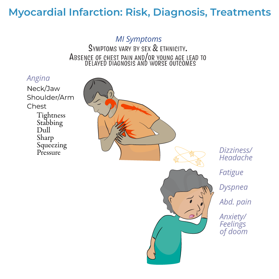
2. Presentation patterns:
- Prodromal symptoms: Days, weeks, or months prior to the acute event
- Acute symptoms: Experienced at the time of the event
- Silent MI: No noticeable symptoms
- Gastrointestinal issues (nausea, vomiting, indigestion)
- Extreme fatigue, exhaustion, or sleep disturbances
- Headaches, dizziness, lightheadedness
- Shortness of breath (dyspnea)
- Anxiety or sense of impending doom
Diagnostic Approach
1. ECG assessment:
- Should be administered as soon as possible when MI is suspected
- Re-administered frequently to observe the evolution of the infarction
- Distinguishes between ST-segment elevated (STEMI) or Non-ST elevated (NSTEMI) myocardial infarctions
- Q-wave abnormalities may indicate size/location of current MI, or may indicate a prior MI
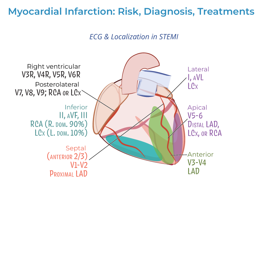
- Lateral infarction: Leads I and aVL; left circumflex artery
- Apical infarction: Leads V5 and V6; left circumflex or right coronary arteries
- Anterior infarction: Leads V3 and V4; left anterior descending artery
- Anteroseptal infarction: Leads V1 and V2; proximal left anterior descending artery
- Inferior infarction: Leads II, aVF, and III; right coronary artery or left circumflex artery (in ~10% with left dominance)
- Right ventricular infarction: Requires additional leads V3R through V6R
- Posterolateral infarction: Requires additional leads V7-V9; right coronary or left circumflex artery
- Cardiac troponin is key to diagnosis
- Help distinguish between NSTEMI and unstable angina (only NSTEMI shows troponin elevation)
- Both cardiac troponin I and CK-MB peak within 24 hours of MI and fall to normal levels over time
Treatment Strategy
1. Time-dependent approach: Treatment should begin as soon as possible, ideally before hospital arrival, to reduce myocardial necrosis.
2. Pre-hospital treatment:
- Oxygen administration when oxygen saturation is less than 90%
- Aspirin for antiplatelet effects
- Nitrates for chest pain (morphine if nitrates ineffective)
- STEMI: Emergency PCI recommended; if unavailable, immediate fibrinolytic therapy
- NSTEMI: Unstable/complicated cases require immediate PCI/CABG; uncomplicated cases may wait longer
- Fibrinolytics: Generally not recommended for NSTEMI (risks outweigh benefits)
- Antiplatelets: Aspirin, clopidogrel, or others
- Anticoagulation: Unfractionated or low molecular weight heparin
- Anti-ischemic: Beta-blockers or calcium-channel blockers
- Plaque stabilization: Statins, ACE-inhibitors
- --
HIGH YIELD
Clinical Recognition Pearls
1. Atypical presentation recognition: Absence of chest pain should not exclude MI from differential, especially in women, elderly, and diabetics.
2. Prodromal symptom importance: Symptoms days to months before acute MI may include fatigue, sleep disturbances, or vague discomfort.
3. Psychological manifestations: Anxiety or sense of impending doom may be harbingers of MI rather than primary psychiatric symptoms.
4. Young patient awareness: Young patients often experience missed or delayed diagnosis due to low clinical suspicion.
5. Diagnostic barriers: Unawareness is a significant obstacle to prevention and treatment—patient education is crucial.
ECG Interpretation Essentials
1. Serial assessment: ECGs should be repeated frequently to observe evolution of infarction patterns.
2. STEMI vs. NSTEMI distinction: Critical for determining appropriate reperfusion strategy.
3. Lead interpretation principles: Different lead changes indicate specific coronary artery territories:
- Anterior (V3-V4): Left anterior descending artery
- Anteroseptal (V1-V2): Proximal left anterior descending artery
- Lateral (I, aVL): Left circumflex artery
- Inferior (II, III, aVF): Right coronary artery (or less frequently left circumflex)
Biomarker Utilization
1. Troponin primacy: Cardiac troponin is key to diagnosis of myocardial infarction.
2. NSTEMI vs. unstable angina differentiation: Only NSTEMI shows rising/falling troponin levels.
3. Temporal pattern: Both cardiac troponin I and CK-MB peak within 24 hours of MI.
4. Serial sampling value: More valuable than single determinations for diagnosis.
5. Integrated interpretation: Always interpret biomarkers in context of clinical presentation and ECG findings.
Treatment Decision-Making
1. Reperfusion timing: "Time is myocardium"—earlier treatment leads to better outcomes.
2. STEMI management: Emergency PCI preferred; if unavailable, immediate fibrinolytic therapy.
3. NSTEMI approach: Risk stratification guides timing of intervention—unstable patients need immediate care.
4. Therapeutic pathway: Comprehensive approach with antiplatelets, anticoagulants, beta-blockers, statins, and ACE inhibitors.
5. Pre-hospital initiation: Early oxygen (when indicated), aspirin, and nitrates can limit infarct size.
Special Population Considerations
1. Women: Higher mortality rates, more atypical presentations, first MI approximately 10 years later than men.
2. Racial/ethnic disparities: Black males have highest incidence after age 35, followed by Black females.
3. Mortality risk: Young and/or minority women have particularly high mortality despite overall declining rates.
4. Age considerations: Young patients often experience missed diagnosis due to low clinical suspicion.
5. Health literacy impact: Many patients, especially women, lack knowledge about risk factors and symptoms—education is essential.
- --
Beyond the Tutorial
Risk Stratification Tools
1. TIMI Risk Score: Predicts 14-day outcomes in ACS patients.
2. GRACE Risk Score: Predicts in-hospital and 6-month mortality.
3. CRUSADE Score: Assesses bleeding risk in ACS patients.
4. DAPT Score: Guides duration of dual antiplatelet therapy after PCI.
5. PRECISE-DAPT Score: Assesses bleeding risk to inform DAPT duration.
Evidence-Based Pharmacotherapy
1. P2Y12 inhibitor selection: Clopidogrel vs. ticagrelor vs. prasugrel based on clinical factors and comorbidities.
2. Anticoagulation strategies: UFH, LMWH, fondaparinux, or bivalirudin based on clinical context.
3. Beta-blocker optimization: Timing, patient selection, and contraindications.
4. High-intensity statin therapy: Specific agents and dosing for post-MI patients.
5. ACE-I/ARB selection: Timing of initiation and patient-specific considerations.
Management of Complications
1. Cardiogenic shock: Early recognition, hemodynamic support options, and revascularization priorities.
2. Mechanical complications: Diagnosis and management of papillary muscle rupture, ventricular septal defect, free wall rupture.
3. Post-MI arrhythmias: Acute management and long-term risk stratification.
4. Right ventricular infarction: Special management considerations including volume loading.
5. Post-infarction pericarditis: Early vs. late (Dressler syndrome) presentations and management.
Secondary Prevention
1. Cardiac rehabilitation: Components, benefits, and appropriate referral.
2. Target goals: LDL <70 mg/dL, BP <130/80 mmHg, HbA1c <7%, smoking cessation.
3. Medication adherence strategies: Improving compliance with life-saving therapies.
4. Return to activities: Evidence-based guidance for driving, sexual activity, exercise, and work.
5. Depression screening: Associated with worse outcomes if untreated.
Quality and Systems-Based Practice
1. Core quality metrics: Door-to-ECG time, door-to-needle time, door-to-balloon time.
2. Regional STEMI systems: Network organization and transfer protocols.
3. Appropriate Use Criteria: For diagnostic and interventional procedures.
4. Discharge planning: Medication reconciliation, follow-up arrangements, and transitions of care.
5. Healthcare disparities: Recognition and strategies to address gaps in MI care and outcomes.