
ABIM - Myocardial Infarction
Start your One-Week Free Trial
Already subscribed? Log in »
Here are key facts for American Board of Internal Medicine (ABIM) Examination from the Myocardial Infarctions: Diagnosis & Treatment tutorial, focusing on clinical management and treatment decision-making that are essential for board certification. See the tutorial notes for further details and relevant links.
 3. Cardiac biomarkers:
 Below is information not explicitly contained within the tutorial but important for the American Board of Internal Medicine Examination.
- --
VITAL FOR ABIM
Epidemiology & Disparities in Care
1. Evolving trends: Incidence of myocardial infarctions is declining in high-income countries but rising in middle- and low-income countries.
2. Demographic variations: Incidence after age 35, from highest to lowest: Black males > Black females > White males > White females.
3. Gender differences: MI occurs approximately 10 years earlier in men than women, possibly related to risk factors like smoking and hyperlipidemia.
4. Mortality patterns: Although overall mortality has declined, rates remain higher in women than men, especially for young and minority women.
5. Disease progression: Myocardial infarction is an important cause of heart failure, which is itself a significant cause of death.
Risk Assessment & Prevention
1. Major modifiable risk factors: Dyslipidemia, diabetes mellitus, hypertension, smoking (including e-cigarettes), obesity, psychosocial stress, alcohol consumption, poor diet (low in fruits and vegetables).
2. Patient awareness challenges: Many patients, especially women, are unaware of risk factors and symptoms—a significant obstacle to prevention and treatment.
3. Pre-infarction symptoms: Prodromal symptoms may occur days, weeks, or even months prior to the actual heart attack.
4. Risk factor management: Long-term treatment focuses on reducing risk factors through improved diet, exercise, and medications for hypertension and hyperlipidemia.
5. Special populations: Understanding demographic-specific clinical presentations improves risk assessment and management.
Clinical Presentation & Evaluation
1. Diagnostic definition: Myocardial infarction is defined as myocardial injury with ischemia.
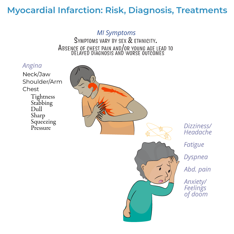
2. Presentation spectrum:
- Typical symptoms: Chest pain/angina (dull, sharp, squeezing, pressure, or discomfort) radiating to arms, neck, jaw, or back
- Atypical presentations: Common in women, elderly, and diabetics
- Silent MI: No noticeable symptoms
Diagnostic Approach
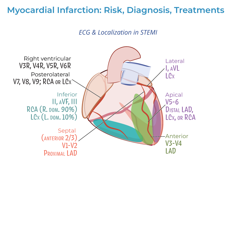
1. ECG interpretation:
- Should be obtained as soon as possible when MI is suspected
- Distinguishes between STEMI and NSTEMI, which influences treatment strategies
- Should be repeated frequently to observe the evolution of the infarction
- Q-wave abnormalities may indicate size/location of current MI or evidence of prior MI
- Lateral infarction: Leads I and aVL; left circumflex artery
- Apical infarction: Leads V5 and V6; left circumflex or right coronary arteries
- Anterior infarction: Leads V3 and V4; left anterior descending artery
- Anterior septal infarction: Leads V1 and V2; proximal left anterior descending artery
- Inferior infarction: Leads II, aVF, and III; right coronary artery or left circumflex artery
- Right ventricular infarction: Requires additional leads V3R through V6R
- Posterolateral infarction: Requires additional leads V7-V9; right coronary or left circumflex artery
3. Cardiac biomarkers:
- Essential for diagnosis, especially cardiac troponin
- Help distinguish between NSTEMI and unstable angina
- Both troponin I and CK-MB peak within 24 hours of the MI and fall to normal levels over time
Treatment Approach
1. Timing principles: Treatment should begin as soon as possible, ideally even before hospital arrival, to reduce the extent of myocardial necrosis.
2. Pre-hospital management:
- Oxygen administration when oxygen saturation is less than 90%
- Aspirin for antiplatelet effects
- Nitrates for chest pain (morphine is an option if nitrates are ineffective)
- Vary by severity of infarction
- Include percutaneous coronary intervention (angioplasty), coronary bypass grafting, or fibrinolytic drugs
- STEMI patients should receive emergency PCI; if unavailable, fibrinolytic drugs must be given as soon as possible
- Unstable, complicated NSTEMI often requires immediate PCI or CABG
- Uncomplicated NSTEMI patients may wait longer, and revascularization may not be necessary
- Fibrinolytic drugs generally not recommended for NSTEMI (risks outweigh benefits)
- Antiplatelets (aspirin, clopidogrel, or others)
- Anticoagulation drugs (unfractionated or low molecular weight heparin)
- Beta-blockers (or calcium-channel blockers)
- Statins
- ACE-inhibitors
- --
HIGH YIELD
Detailed Clinical Presentations
1. Symptom variability: Chest pain or angina is variably described as dull, sharp, squeezing, pressure, or simply discomfort—understanding this spectrum improves recognition.
2. Pain radiation patterns: May radiate from the chest to arms, neck, jaw, or back—radiation patterns can help confirm diagnosis.
3. Non-chest pain presentations: Not all patients experience angina; absence increases risk of missed diagnosis, especially in women and elderly.
4. Prodromal symptoms: May occur days, weeks, or even months before the acute event—recognition can enable earlier intervention.
5. Psychological manifestations: Anxiety or sense of impending doom may precede or accompany MI—should not be dismissed as purely psychiatric.
ECG Interpretation Pearls
1. Serial assessment: ECGs should be repeated frequently to observe the evolution of infarction patterns.
2. STEMI vs. NSTEMI distinction: Critical for determining appropriate reperfusion strategy.
3. Localization significance: Different lead changes indicate specific coronary artery territories:
- Anterior (V3-V4): Left anterior descending artery
- Anteroseptal (V1-V2): Proximal left anterior descending artery
- Lateral (I, aVL): Left circumflex artery
- Apical (V5-V6): Left circumflex or right coronary arteries
- Inferior (II, III, aVF): Right coronary artery (or less frequently left circumflex)
Biomarker Interpretation
1. Diagnostic utility: Cardiac biomarkers, especially troponin, are key to diagnosing myocardial infarction.
2. Differential diagnosis: Biomarker values help distinguish between NSTEMI and unstable angina—only NSTEMI shows rising/falling troponin.
3. Temporal patterns: Both cardiac troponin I and CK-MB peak within 24 hours of MI and gradually return to normal.
4. Sampling protocols: Serial measurements provide more diagnostic value than single determinations.
5. Integration with clinical findings: Biomarkers should be interpreted in the context of symptoms and ECG findings.
Reperfusion Decision-Making
1. STEMI management: Emergency PCI recommended; if unavailable, fibrinolytic drugs must be given as soon as possible.
2. NSTEMI approach: Timing of intervention based on risk stratification:
- Unstable/complicated cases: Immediate PCI/CABG
- Uncomplicated cases: May wait longer; revascularization may not be necessary
Special Populations Considerations
1. Gender differences: Women experience MI approximately 10 years later than men, often with atypical symptoms, and have higher mortality rates.
2. Racial/ethnic variations: Significant differences in MI incidence across populations, with Black males having highest incidence after age 35.
3. Young patients: Absence of chest pain and young age often leads to missed diagnosis—maintain high index of suspicion.
4. Awareness disparities: Many patients, especially women, are unaware of risk factors and symptoms—patient education is crucial.
5. Risk factor clustering: Combinations of risk factors like smoking, dyslipidemia, and diabetes significantly increase MI risk.
- --
Beyond the Tutorial
Advanced Risk Stratification
1. TIMI Risk Score: Predicts 14-day and 1-year mortality risk in STEMI/NSTEMI patients.
2. GRACE Risk Score: Predicts in-hospital and 6-month mortality for ACS patients.
3. HEART Score: Stratifies chest pain patients in the emergency department for early discharge consideration.
4. Bleeding risk assessment: CRUSADE score for NSTEMI and HAS-BLED for anticoagulated patients.
5. Long-term risk prediction: ASCVD risk calculator for secondary prevention intensity.
Contemporary Management Strategies
1. Pharmaco-invasive approach: Pre-hospital fibrinolysis followed by early angiography in certain settings.
2. P2Y12 inhibitor selection: Clopidogrel vs. ticagrelor vs. prasugrel based on patient characteristics.
3. Anticoagulant options: UFH, LMWH, fondaparinux, or bivalirudin based on clinical scenario.
4. Complete vs. culprit-only revascularization: Strategies for multivessel disease in STEMI.
5. Optimal timing of non-culprit lesion intervention: Immediate vs. staged approach.
Complications Management
1. Mechanical complications: Recognition and management of papillary muscle rupture, ventricular septal defect, and free wall rupture.
2. Cardiogenic shock: Early recognition, hemodynamic support, and revascularization strategies.
3. Post-MI arrhythmias: Acute management and long-term risk stratification.
4. Right ventricular infarction: Special management considerations including volume resuscitation.
5. Post-infarction pericarditis: Differentiation from reinfarction and appropriate therapy.
Secondary Prevention and Follow-up
1. Optimal medical therapy: Evidence-based medication regimens and dosing.
2. Cardiac rehabilitation: Structured programs to improve outcomes and reduce readmissions.
3. Implantable device considerations: ICD and CRT evaluations post-MI.
4. Return to activities: Evidence-based guidance for driving, physical activity, and work.
5. Long-term monitoring: Appropriate use of testing for residual ischemia and ventricular function assessment.
Quality and Systems-Based Practice
1. Core quality metrics: Door-to-ECG time, door-to-needle time, door-to-balloon time.
2. Regional STEMI systems: Network organization and transfer protocols.
3. Discharge planning: Medication reconciliation, follow-up arrangements, and transition of care.
4. Readmission reduction: Evidence-based strategies to prevent 30-day readmissions.
5. Healthcare disparities: Recognition and addressing gaps in MI care and outcomes.