

Start your One-Week Free Trial
Already subscribed? Log in »

Urinary Bladder
The ureters and urinary bladder are retroperitoneal structures in the abdomen and pelvis (some consider the urinary bladder to be subperitoneal or infraperitoneal).
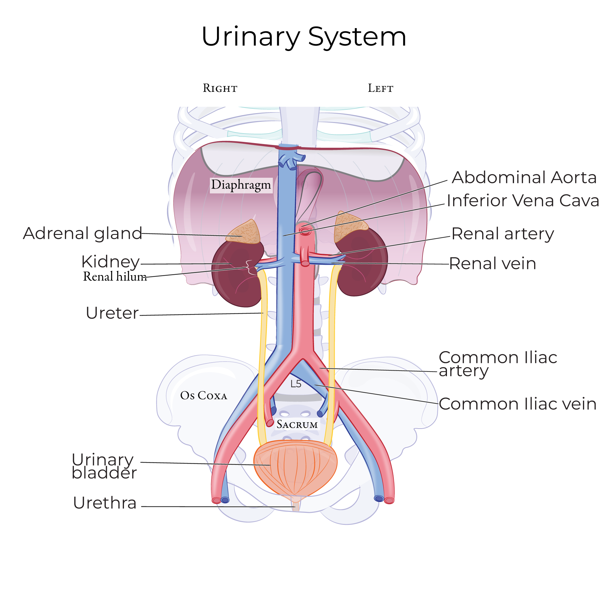
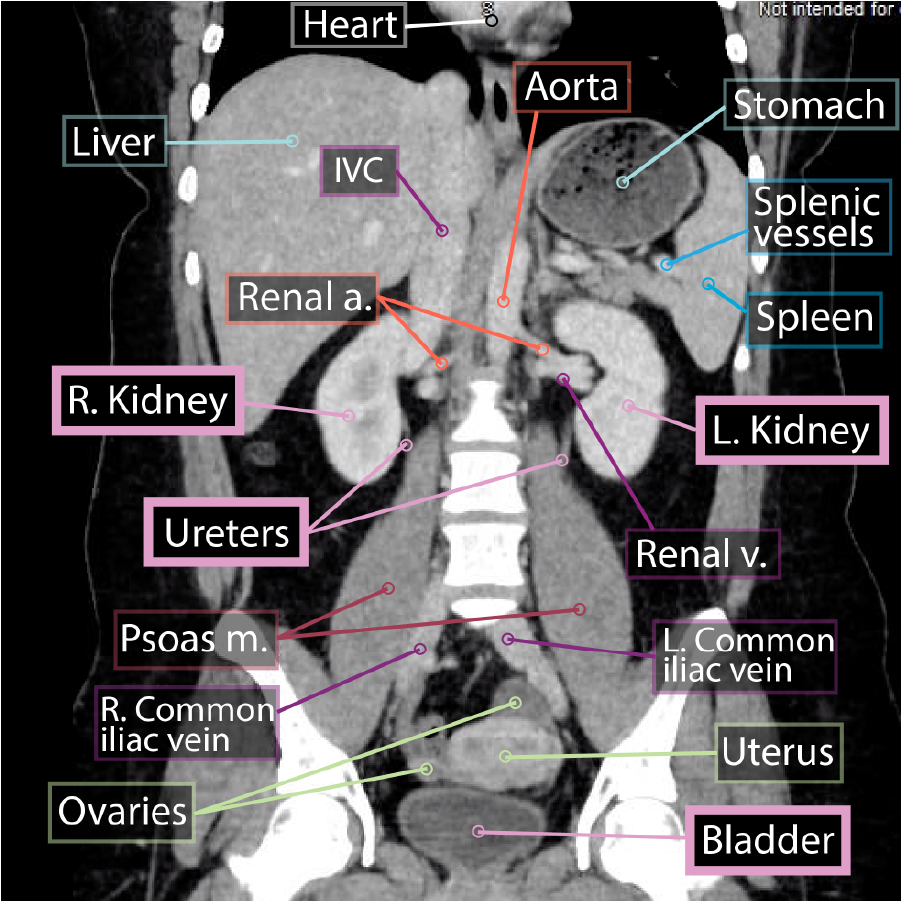 The kidneys lie on the posterior abdominal wall; these organs filter the blood and produce urine.
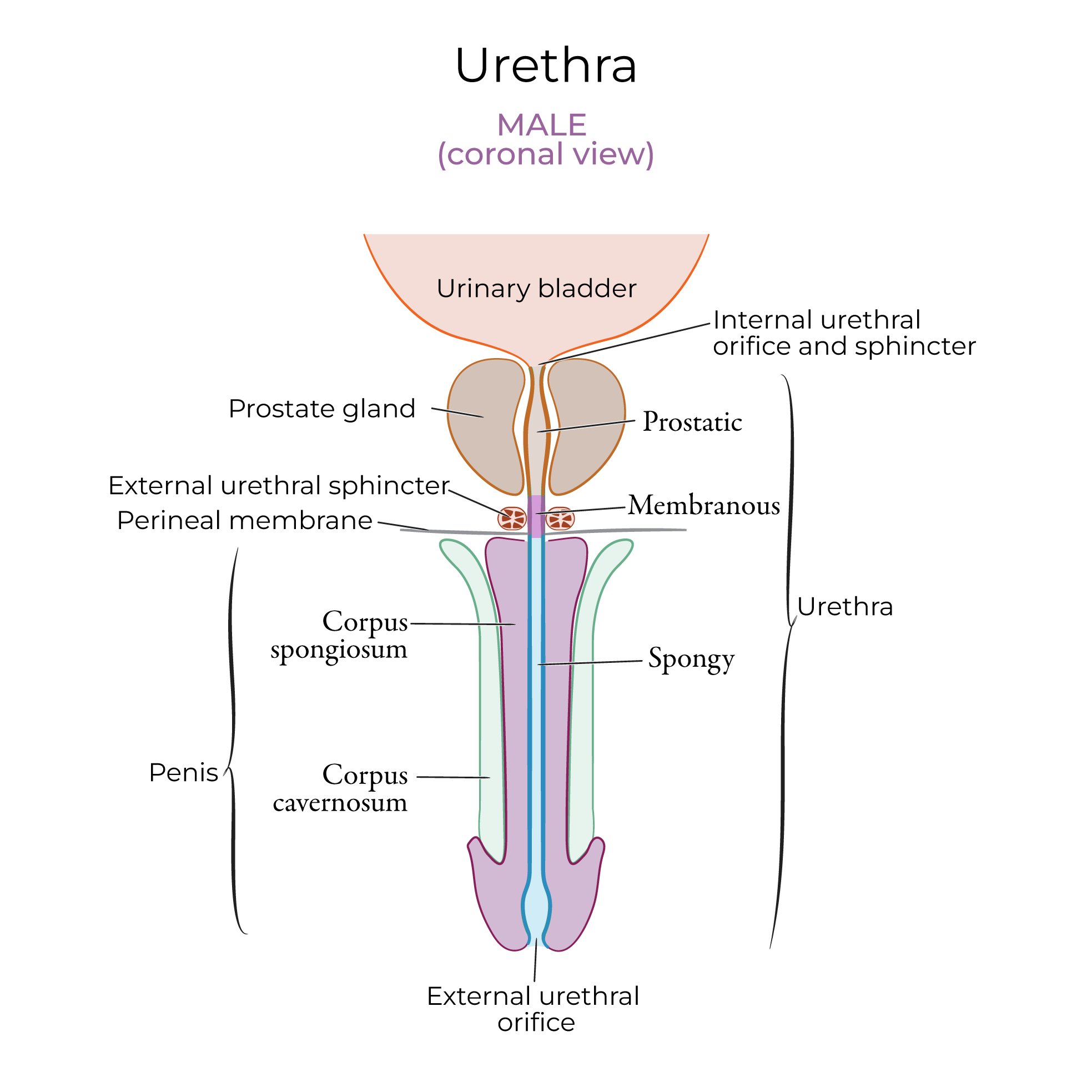
 In males, the urethra travels the length of the penis, so the external urethral orifice is located at the tip of the penis.
 Urinary incontinence is defined as the loss of bladder control with unintentional voiding. Types of incontinence include stress incontinence, which is the result of weak urinary sphincters and urge incontinence, which is caused by an overactive detrusor muscle.
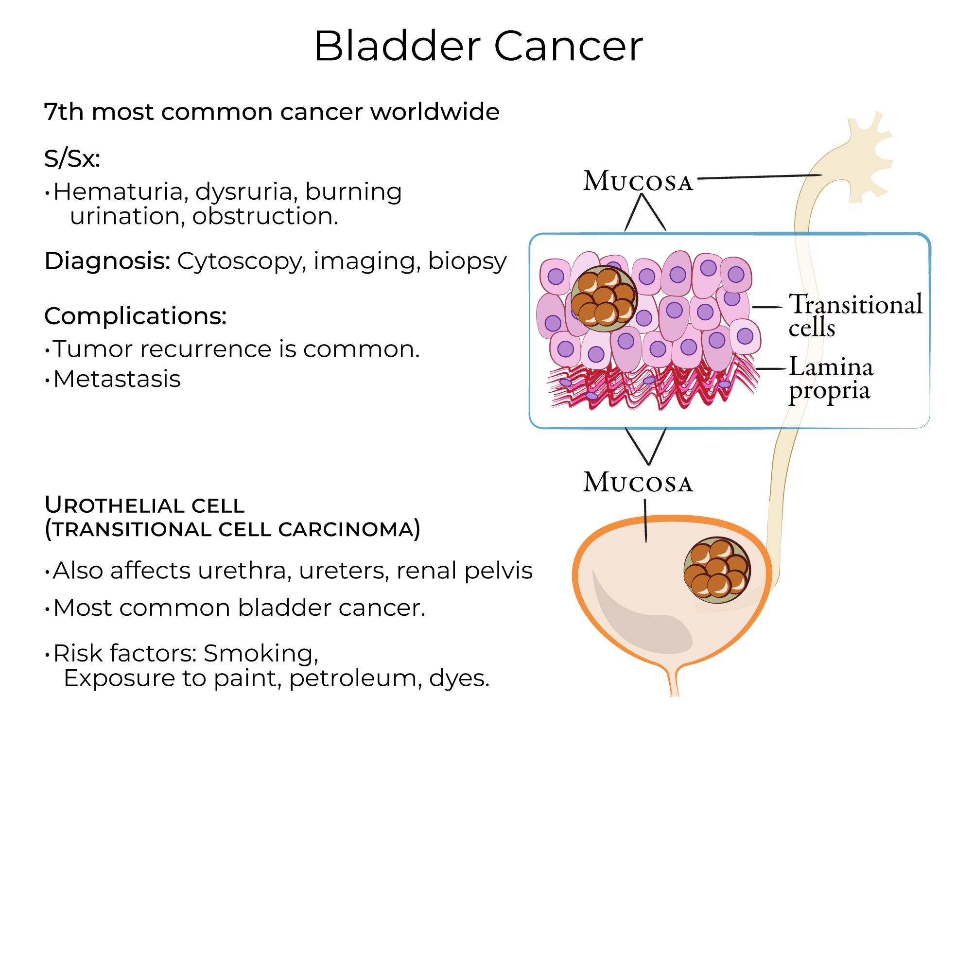
Bladder cancer is a common cancers worldwide; urothelial cell carcinoma, which arises in the transitional epithelium, accounts for approximately 90% of cases. Be aware that this cancer can occur anywhere along the urinary tract.
Urinary incontinence is defined as the loss of bladder control with unintentional voiding. Types of incontinence include stress incontinence, which is the result of weak urinary sphincters and urge incontinence, which is caused by an overactive detrusor muscle.
Bladder cancer is a common cancers worldwide; urothelial cell carcinoma, which arises in the transitional epithelium, accounts for approximately 90% of cases. Be aware that this cancer can occur anywhere along the urinary tract.
 Urinary tract obstruction
The kidneys lie on the posterior abdominal wall; these organs filter the blood and produce urine.
The Urinary Bladder
The adventitia is its outermost layer.
The muscularis layer comprises the detrusor muscle. The detrusor comprises three layers of smooth muscle that is relaxed when the bladder is storing urine, and contracts to expel urine through the urethra.
Deep to detrusor, show the submucosa, which is a layer of supportive connective tissues.
The mucosa comprises lamina propria and transitional epithelia.
Transitional epithelium lines the ureters, urinary bladder, and the proximal urethra; this specialized epithelium comprises cells that change shape to accommodate changes in urine volume.
Transitional epithelium comprises stratified layers of cells that, when relaxed, are cuboidal. Umbrella cells within the transitional epithelium can flatten to expand and accommodate changes in urine volume. Note that these cells are often binucleated, which helps us to identify them under the microscope.
The mucosa forms rugae on the internal surface of the urinary bladder; these folds expand to accommodate increased urine volume.
The ureters open on the posterior wall of the bladder; the internal urethral orifice and sphincter are at its inferior apex.
The opening is the exit for urine, and the sphincter comprises smooth muscle that regulates urine passage through the orifice.
The trigone is the triangular area bound by the lateral ureter openings and the inferior internal urethral orifice; this is a region of the bladder wall that acts as a funnel to direct urine through the neck of the bladder and into the urethra when the bladder contracts. Embryologically, the trigone is derived from the ends of the mesonephric ducts.
The external urethral sphincter is located in the deep perineal pouch. Be aware that some texts describe the external urethral sphincter within the urogenital diaphragm, which is an older anatomical term.
The external urethral sphincter comprises skeletal muscle and is what we use to “hold” our urine consciously.
The external urethral orifice is were urine exits the body.
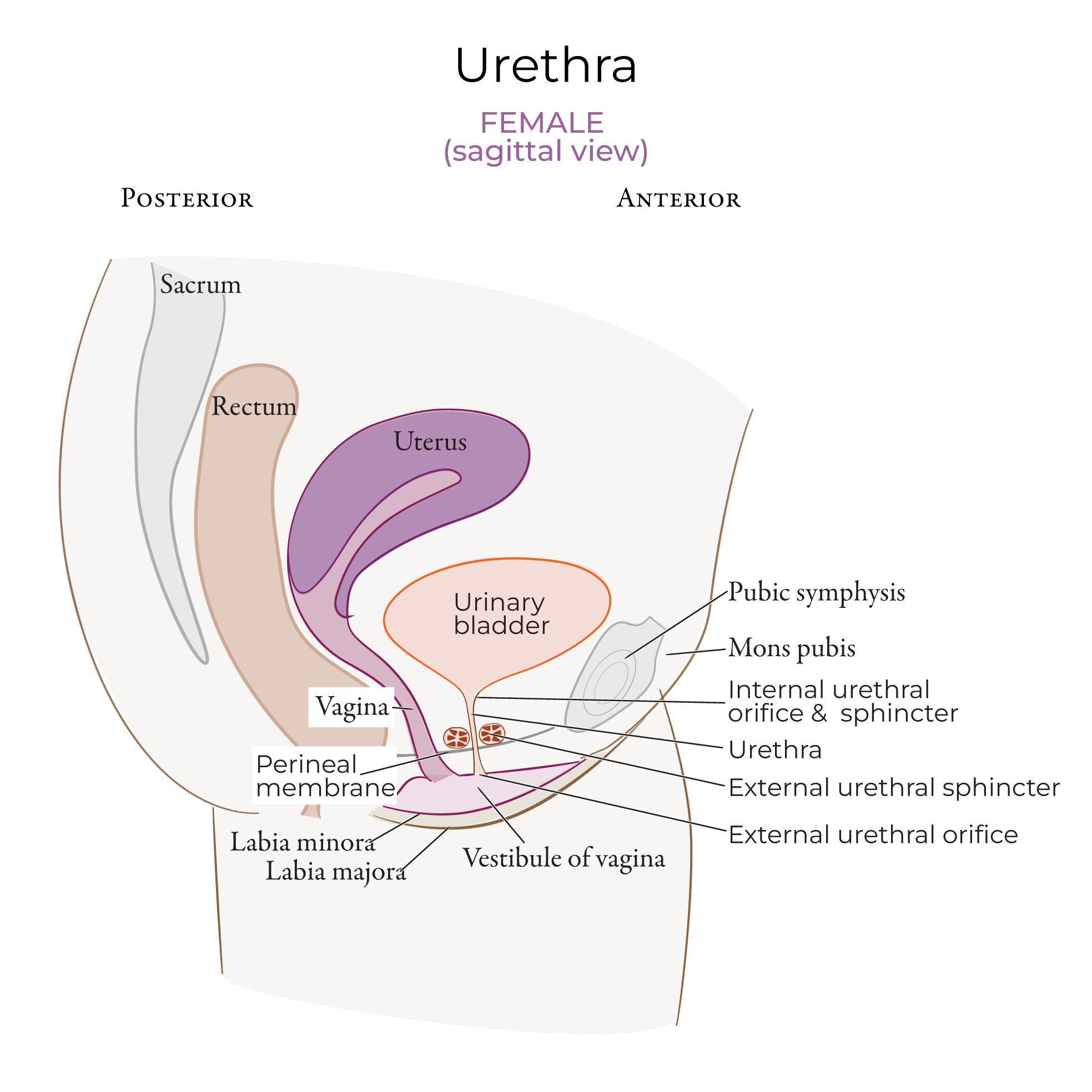
In females, the urethra is 3-5 centimeters long, and the external urethral orifice is located within the perineum.
In males, the urethra travels the length of the penis, so the external urethral orifice is located at the tip of the penis.
Clinical Correlations
Urinary tract obstruction