

Start your One-Week Free Trial
Already subscribed? Log in »

Posterior Hip Muscles
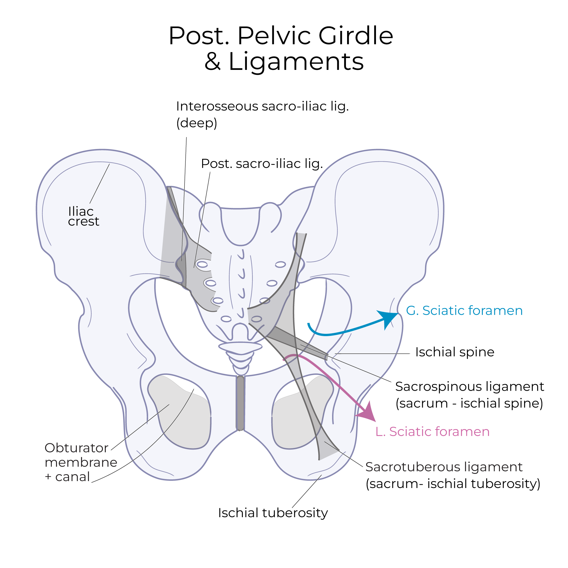
Overview of the Posterior Pelvic Girdle & Ligaments
- Iliac crest, superiorly
- Ischial spine
- Ischial tuberosity, inferiorly
- Gluteal lines serve as attachment points for the gluteal muscles: Posterior, Anterior, and Inferior.
Key Connective Tissues & Ligaments:
Obturator membrane
- Covers the obturator foramen.
- The obturator canal is a small opening in the membrane. The obturator nerve travels through this canal from the pelvic cavity to the anterior/medial thigh.
Interosseous sacro-iliac ligament (deep)
- As its name suggests, it secures the sacrum and ilium.
Posterior sacro-iliac ligament
- A larger, more superficial ligament with fibers that run in multiple directions to secure the pelvic bones.
Sacro-spinous ligament
- Runs from the sacrum to the ischial spine.
Sacro-tuberous ligament
- Runs from the sacrum to the ischial tuberosity.
Sciatic Notch, Foramina & Contents
- The interosseous sacroiliac and posterior sacroiliac ligaments pass over the sciatic notch, converting it into foramina.
- The greater sciatic foramen, superior to the sacrospinous ligament.
- The lesser sciatic foramen, inferior to the ligament.
- These foramina provide entry and exit points to the pelvis and perineum.
Greater sciatic foramen
- Divided into superior and inferior portions by the piriformis muscle.
- Above the piriformis: the superior gluteal vessels and nerve travel to the glutes.
- Below the piriformis, several neurovascular structures pass: sciatic nerve, inferior gluteal vessels and nerve, internal pudendal vessels and nerve, posterior femoral cutaneous nerve and the nerve to obturator internus & superior gemellus muscles, and, the nerve to quadratus femoris and inferior gemellus muscles.
Lesser sciatic foramen
- The tendon for obturator internus muscle exits to the greater trochanter, and, the pudendal nerve and vessels pass back from the gluteal region to the perineal region.
Deep Muscles of the Posterior Hip
Quadratus femoris
- Arises from the ischial tuberosity and inserts on the intertrochanteric crest of the femur.
- As its name suggests, this is a rectangular ("quadratic") muscle; "femoris" refers to the femur – be careful not to confuse this muscle with rectus femoris, a long muscle in the anterior thigh.
Inferior gemellus
- Arises from the ischial tuberosity and inserts on the greater trochanter of the femur.
Obturator internus
- Arises from the obturator membrane and the ischiopubic rami and inserts on the greater trochanter via a thin tendon.
Superior gemellus
- Arises from the ischial spine and inserts on the greater trochanter.

Piriformis
- A flat, broader muscle that arises from the anterior surface of the sacrum and passes through the greater sciatic foramen to attach on the greater trochanter of the femur.
 For context, we show gluteus minimus to see its relationship to these rotators.
Superficial Muscles of the Posterior Hip
Gluteus minimus
- The smallest and deepest of the gluteal muscles.
- Arises from the ilium, between the anterior and inferior gluteal lines, and inserts on the greater trochanter of the femur.
Gluteus medius:
- Arises superior to gluteus minimus, between the anterior and posterior gluteal lines, and also inserts on the greater trochanter of the femur.
Notice that the fibers of gluteus medius and minimus run in similar directions; this helps us to remember that both act as abductors and medial rotators of the thigh.
Tensor fasciae latae
- A small muscle that arises from the iliac crest and anterior superior iliac spine; this muscle is also visible in anterior and lateral views.
- The fascia that covers this muscle extends inferiorly as the iliotibial band, which attaches to the tibia (not shown); thus, the iliotibial band, aka, the IT band or tract, stabilizes the knee.
Gluteus maximus
- The largest muscle in the hip
- Arises from the posterior ilium and sacrum (specifically, from the sacro-tuberous ligament) and extends inferiorly and laterally; the deeper fibers of this thick muscle attach to the gluteal tuberosity of the femur, whereas the more superficial and anterior fibers attach along the iliotibial band.
Sciatic Nerve & Piriformis Syndrome
Posterior Hip Innervation
- Piriformis is innervated by the anterior rami of S1 and S2.
- Superior gemellus and obturator internus are innervated by the nerve to obturator internus; inferior gemellus and quadratus femoris are innervated by the nerve to quadratus femoris.
- Tensor fascia latae and the two deeper gluteal muscles are innervated by the superior gluteal nerve, and gluteus maximus is innervated by the inferior gluteal nerve.