

Start your One-Week Free Trial
Already subscribed? Log in »


Large Intestine
Key Features
- Three longitudinal ribbon-like bands of muscle fibers that travel the length of the large intestine. The taeniae coli represent the muscularis tunic of the large intestine.
- They act like an elastic band that pulls on the large intestine and causes it to bunch and form haustra.
- Haustra
- Pouch-like structures.
- Epiploic appendages (aka, omental appendages)
- Small fat-filled sacs, attach to the tenaie coli.
Key Functions
- Receives undigested materials from the small intestine.
- Absorbs water and ions from the undigested materials, which converts the remaining materials to feces (the small intestine is the primary place of nutrient absorption).
- Stores and expels feces.
Subdivisions of the large intestine
- Cecum (appendix attaches, here)
- Ascending colon
- Transverse colon
- Descending colon
- Sigmoid colon
- Rectum
- Anal canal, which opens to external environment via the anus.
- External and internal anal sphincters regulate passage of feces.
- External anal sphincter comprises voluntary skeletal muscle
- Internal anal sphincter comprises involuntary smooth muscle
Key Landmarks:
- Right colic flexure (aka, hepatic flexure)
- Left colic flexure (aka, splenic flexure)
- Distal sigmoid colon and rectum lie within the pelvis.
- Anal canal lies within the perineum, external to the abdominopelvic cavity.
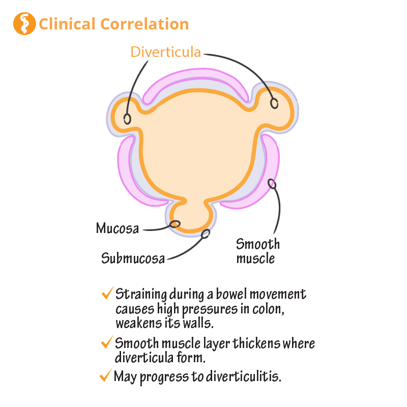
Clinical Correlations
 Inflammatory bowel disease (IBD) refers to chronic inflammation of the GI tract
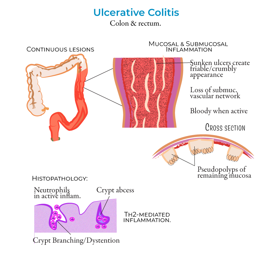
Ulcerative colitis causes continuous ulcers, specifically within the lining of the large intestine.
Inflammatory bowel disease (IBD) refers to chronic inflammation of the GI tract
Ulcerative colitis causes continuous ulcers, specifically within the lining of the large intestine.
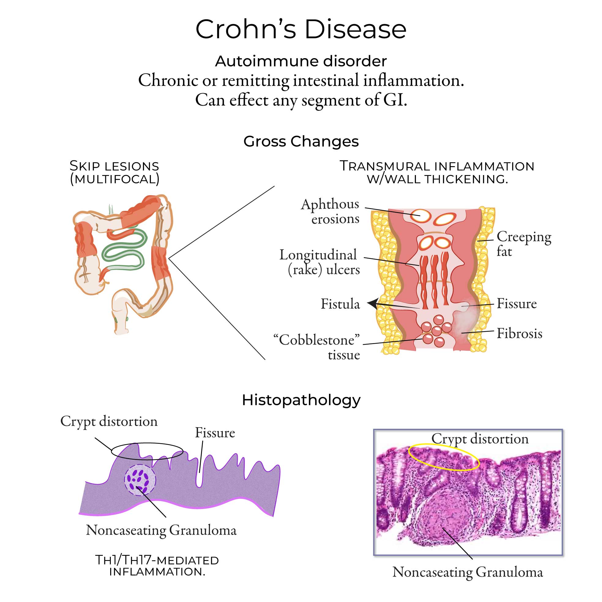
 Crohn's disease is characterized by inflammation that spreads deep into the walls of the GI tract.
CT Scans


 For more, see our Abdominal CT Scan Atlas on the Course Page