

Start your One-Week Free Trial
Already subscribed? Log in »

Heart - External Features
Here we'll learn the external features of the heart.
The heart is roughly triangular in shape; in anatomical position, its apex points slightly towards the left side of the body.
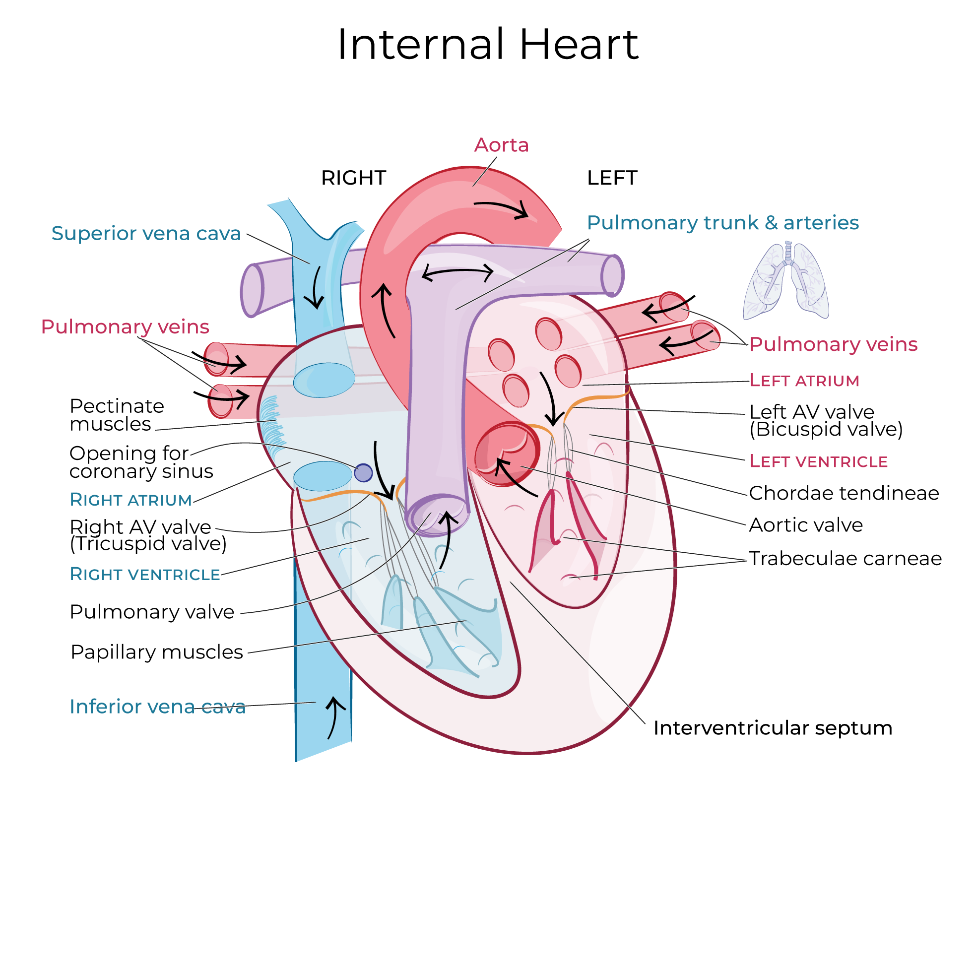
Internally, the heart is divided into four chambers, which we'll approximate on the external surface: the smaller chambers, the atria, are superior and the ventricles are inferior.
 The auricles are ear-like extensions of the atria; when necessary, these pouches can expand to accommodate blood flow.
The coronary sulcus (aka, atrioventricular groove) is a shallow depression inferior to the auricles between the atria and ventricles.
The coronary sulcus provides space for the coronary arteries and the Great cardiac vein; we show that epicardial adipose (fatty) tissue surrounds the vessels.
The auricles are ear-like extensions of the atria; when necessary, these pouches can expand to accommodate blood flow.
The coronary sulcus (aka, atrioventricular groove) is a shallow depression inferior to the auricles between the atria and ventricles.
The coronary sulcus provides space for the coronary arteries and the Great cardiac vein; we show that epicardial adipose (fatty) tissue surrounds the vessels.
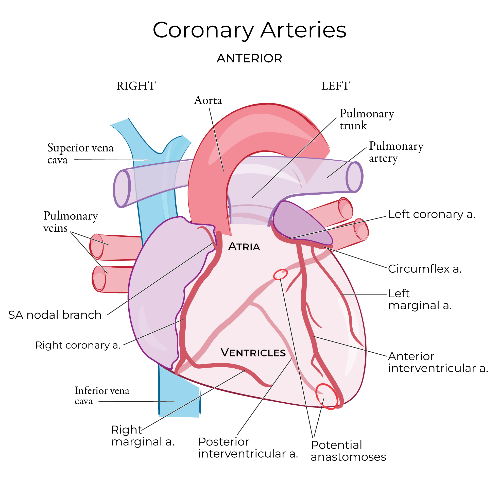 The anterior interventricular sulcus is a shallow depression between the right and left ventricles. The anterior interventricular artery and vein travel within the sulcus, surrounded by fatty tissue.
The base of the heart is the widest part of the heart, and primarily overlies the left atrium.
Posteriorly, the coronary sulcus contains the coronary sinus, small cardiac vein, right coronary artery, and the circumflex branch of the left coronary artery.
The posterior interventricular sulcus contains the middle cardiac vein and the posterior interventricular artery.
The anterior interventricular sulcus is a shallow depression between the right and left ventricles. The anterior interventricular artery and vein travel within the sulcus, surrounded by fatty tissue.
The base of the heart is the widest part of the heart, and primarily overlies the left atrium.
Posteriorly, the coronary sulcus contains the coronary sinus, small cardiac vein, right coronary artery, and the circumflex branch of the left coronary artery.
The posterior interventricular sulcus contains the middle cardiac vein and the posterior interventricular artery.
Anterior Heart
The auricles are ear-like extensions of the atria; when necessary, these pouches can expand to accommodate blood flow.
The coronary sulcus (aka, atrioventricular groove) is a shallow depression inferior to the auricles between the atria and ventricles.
The coronary sulcus provides space for the coronary arteries and the Great cardiac vein; we show that epicardial adipose (fatty) tissue surrounds the vessels.
The anterior interventricular sulcus is a shallow depression between the right and left ventricles. The anterior interventricular artery and vein travel within the sulcus, surrounded by fatty tissue.
The Great Vessels
Review Systemic & Pulmonary Circulation
The pulmonary trunk arises near the left auricle, then splits to form the right and left pulmonary arteries, which carry deoxygenated blood from the heat to the lungs.
The aorta arises at an angle and wraps around the pulmonary trunk; we label the ascending segment and the arch of the aorta. The aorta delivers oxygenated blood from the heart to the rest of the systemic circulatory system.
The superior and inferior vena cavae lie posteriorly; they return deoxygenated blood from the body to the atria of the heart.
The paired right and left pulmonary veins deliver oxygenated blood from the lungs to the posterior aspect of the left atrium.
Posterior Heart
Great vessels
The superior and inferior vena cavae drain into the right atrium, and the pulmonary veins drain into the left atrium.
Blood is delivered from the lungs to the atria via these vessels.
The pulmonary trunk arises from the anterior surface and splits to form the pulmonary arteries.
The aorta arises from the anterior surface and wraps posteriorly over the pulmonary trunk.
The pulmonary arteries and aorta deliver blood from the ventricles to the body.
Notice that the vena cavae and aorta, which are the primary branches of the systemic circulation, extend along the vertical axis of the body.
The pulmonary arteries and veins run more horizontally because they transport blood to and from the lungs, which are situated to the right and left sides of the heart.