

Start your One-Week Free Trial
Already subscribed? Log in »

Arteries - Trunk & Abdominal Walls
Here we'll learn the arteries that supply the walls of the trunk (the thoracic cage and the abdomen).
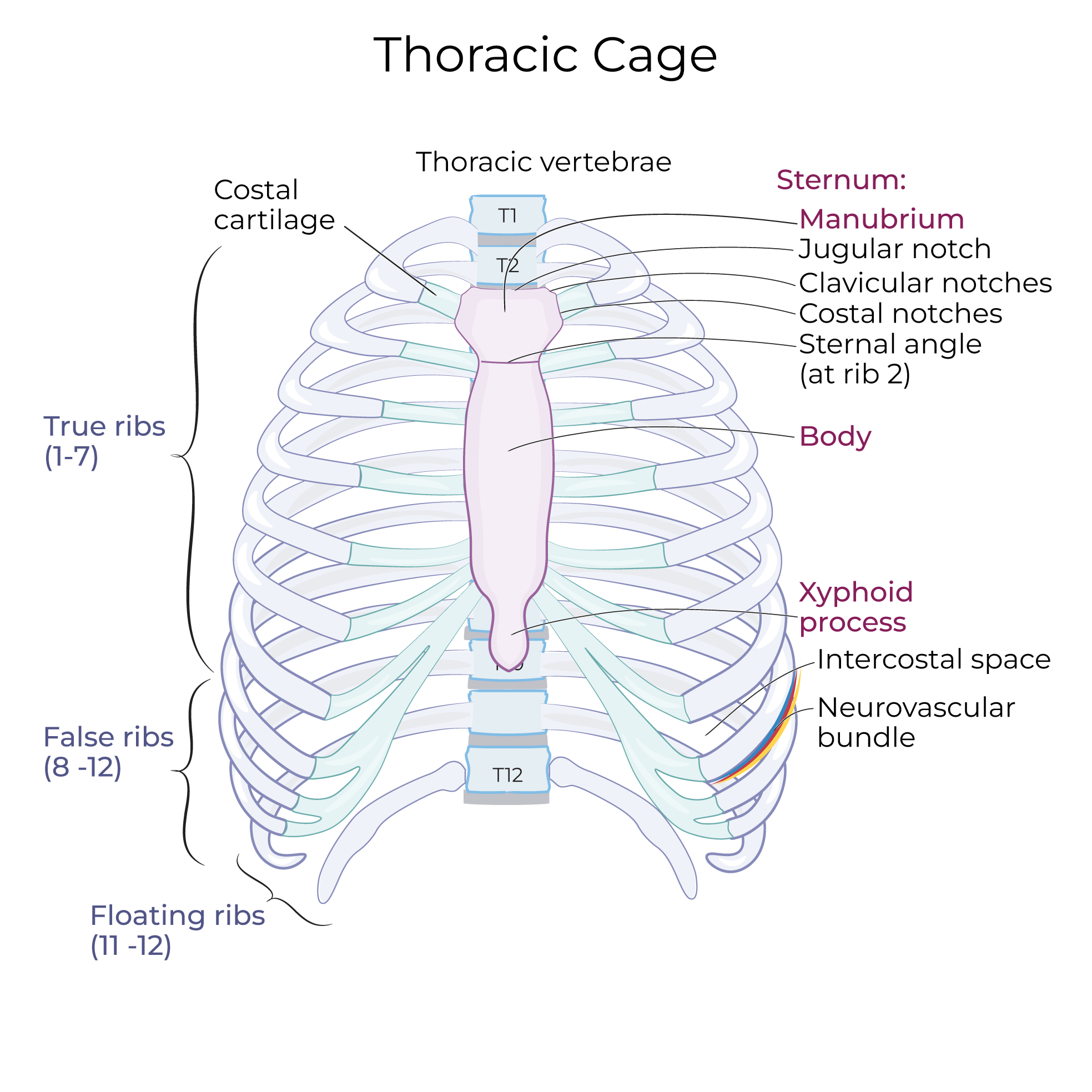
We show the relevant skeletal features: the vertebral column, pelvis, and thoracic cage.
 Be aware that we've simplified the anterior intercostal arteries in our diagram; in reality, they are paired, and two arteries travel within the intercostal spaces.
Simplified Flowchart
- Start with the aorta and the external iliac arteries.
- On the "superficial" side of this flowchart, we show that the aorta gives rise to the subclavian artery, which gives rise to the internal thoracic artery.
- The internal thoracic artery terminates by transitioning to the superior epigastric artery.
- The inferior epigastric artery and the ascending branch of the deep circumflex arteries arise from the external iliac arteries.
- On the "deep" side of this flowchart, show that the subclavian artery gives rise to the costocervical trunk, which gives rise to a branch called the superior intercostal artery.
- The superior intercostal artery gives rise to the first and second posterior intercostal arteries.
- The following arteries arise directly from the aorta: Posterior intercostal arteries 3-11, the subcostal artery, and 4 lumbar arteries.
- The median sacral artery arises from the distal end of the aorta where it bifurcates.
Detailed Diagram
- Start with the aorta, which has ascending, arching, and descending portions.
- The descending aorta bifurcates distally to give rise to the common iliac arteries; these arteries then split to give rise to the internal and external iliac arteries.
- The right and left subclavian arteries: on the right side, the brachiocephalic artery arises from the arch of the aorta and splits to form the subclavian and internal carotid arteries; on the left side, the internal carotid and subclavian arteries arise directly from the aorta.
- On the superficial side of our diagram, show that the internal thoracic artery arises from the subclavian artery and travels along the internal aspect of the anterior thoracic wall.
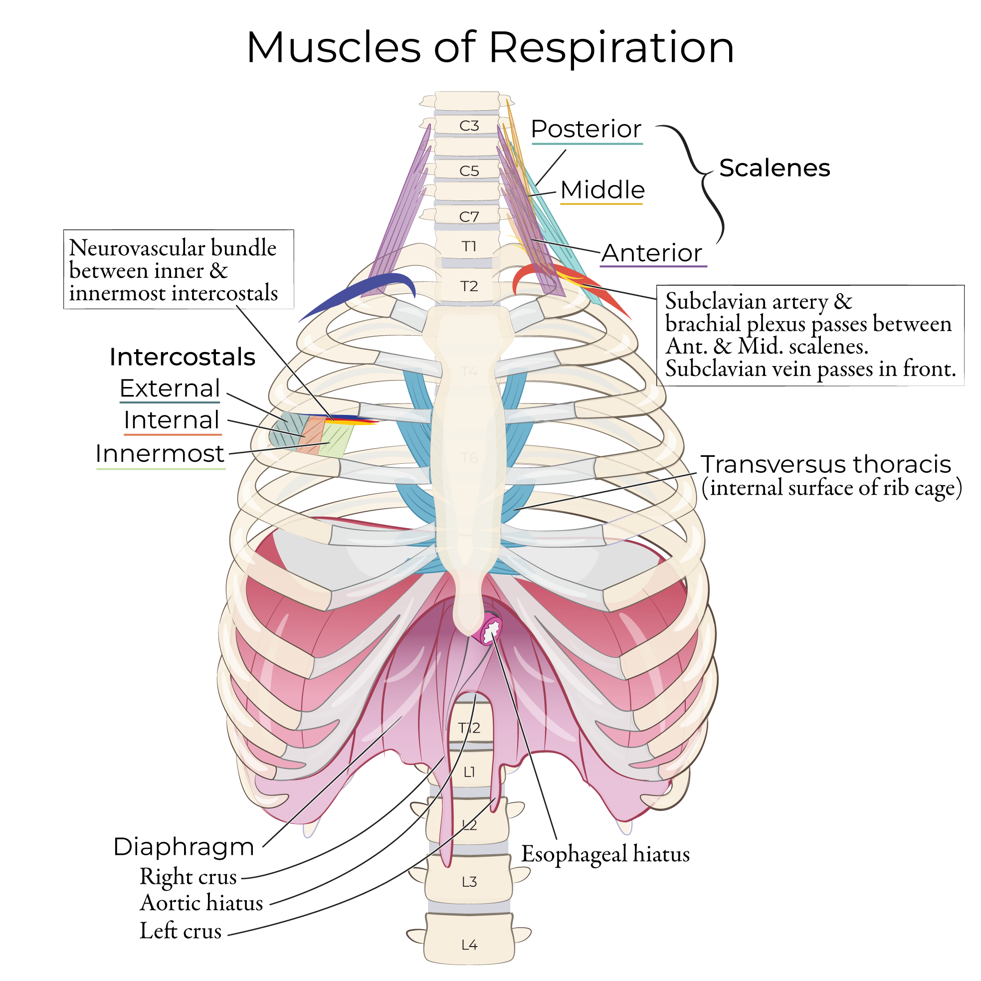
- As it descends through the thorax, the internal thoracic artery gives rise to the first six anterior intercostal arteries, which travel in the intercostal spaces with the intercostal veins and nerves. Specifically, between the innermost and inner intercostal muscles.
Be aware that we've simplified the anterior intercostal arteries in our diagram; in reality, they are paired, and two arteries travel within the intercostal spaces.
- At approximately the 6th intercostal space, the internal thoracic artery branches to give rise to the musculophrenic artery, which gives off anterior intercostal arteries 7-9. As its name suggests, this artery also serves the diaphragm.
- There are no anterior intercostal arteries in the remaining intercostal spaces (but we'll see posterior intercostal arteries in all spaces).
- The internal thoracic artery terminates as the superior epigastric artery.
- The external iliac artery gives rise to the inferior epigastric artery; this artery ascends along the abdominal wall and anastomoses with the superior epigastric artery. Together, these arteries and their branches are the primary blood supply for the anterior abdominal wall.
- The ascending branch of the deep circumflex artery also arises from the external iliac artery. This branch also travels proximally and forms anastomoses with other arteries to supply the abdominal wall.
- The subclavian artery gives rise to the costocervical trunk, which in turn gives off the superior intercostal artery (aka supreme intercostal artery).
- The superior intercostal artery gives rise to the first and second posterior intercostal arteries.
- Posterior intercostal arteries 3-11 arise directly from the descending thoracic aorta.
- The posterior and anterior intercostal arteries anastomose in the intercostal spaces, forming a continuous arterial supply around the ribcage.
- Under rib 12, show that the subcostal artery arises directly from the abdominal aorta; then, move lower, and show the 4 pairs of lumbar arteries.
- The median sacral artery extends from the midline where the aorta bifurcates; this is the only unpaired vessel, be aware that it give off several anastomosing branches, and, occasionally a 5th set of lumbar arteries.