

Start your One-Week Free Trial
Already subscribed? Log in »
Urinary System Anatomy Overview
Here we'll learn an overview of the urinary system, which is located in the abdominal and pelvic cavities.
The kidneys filter blood and produce urine.
The ureters are paired tubes that carry the urine from the kidneys to the urinary bladder.
The urinary bladder is an expandable muscular sac that collects and stores urine; when the urinary bladder contracts, urine is expelled through the urethra, which carries urine to the external environment.
Every day, the kidneys filter 150-200 liters of blood plasma and produce about 2 liters of urine.
Filtration and excretion removes wastes and toxins from the blood and regulates ion and acid/base balance.
By regulating how much water is excreted in the urine, the kidneys play a role in maintaining blood volume and, therefore, pressure.
The kidneys respond to low oxygen levels (hypoxia) by releasing erythropoietin, which stimulates red blood cell production in the bone marrow.
The kidneys also convert vitamin D to its active form; vitamin D is necessary for bone growth.
Anatomical Context:
We show the lower thoracic spine, the lumbar spine, the sacrum and os coxa; we also show the bottom ribs and indicate where the dome-shaped muscular diaphragm marks the superior border of the abdominal cavity; we show the lower rib cage cut open around it.
The internal and external iliac veins converge to form the common iliac veins within the pelvis; the right and left common iliac veins converge to form the inferior vena cava at approximately lumbar vertebra 5.
The Inferior vena cava ascends through the abdominal cavity and exits the abdominal cavity through the caval opening in the diaphragm.
The descending aorta passes through the aortic hiatus of the diaphragm; for context, show the celiac trunk and superior mesenteric artery.
The abdominal aorta splits to form the right and left common iliac arteries, which split again to form the internal and external iliac arteries in the pelvis.
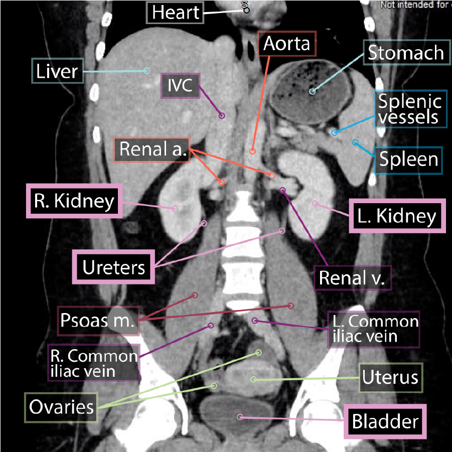 The right and left adrenal, aka suprarenal glands sits on the superior poles of the kidneys.
The hilum is on the medial aspect of the kidney; this is where the renal vessels, nerves, and ureters enter and exit the kidney.
The renal arteries are deeper, and branch directly off the abdominal aorta; notice that the right renal artery travels behind the inferior vena cava.
The renal veins are superficial to the arteries, and drain directly into the inferior vena cava; notice that the left renal vein passes under the superior mesenteric artery.
Innervation is achieved via the renal plexus, which contains sympathetic fibers (from spinal segments T10-12) and parasympathetic fibers (from the vagus nerve).
The ureters descend through the abdominal cavity and into the pelvis; notice that they travel over the iliac vessels before draining urine into the posterolateral walls of the urinary bladder. The ureters are retroperitoneal and are approximately 25 cm long.
The urinary bladder is mostly retroperitoneal (some texts refer to it as subretroperitoneal or infraperitoneal because of its position in the pelvic cavity). It sits immediately posterior to the pubic symphysis of the pelvis; when present, the vagina sites immediately posterior to the urinary bladder and the uterus is superior and posterior.
Urine exits the body via the urethra, which is a thin-walled fibromuscular tube regulated by internal and external sphincters.
The internal sphincter is located in the neck of the bladder at the origin of the urethra; it comprises involuntary smooth muscle and regulates the passage of urine from the urinary bladder to the urethra.
The external sphincter comprises voluntary skeletal muscle fibers derived from the urogenital diaphragm. This is the muscle that we learn to control when we learn to control where and when we void our bladders (toilet training).
Anatomy of micturition (urination).
In females, the urethra is relatively short, 3-5 cm long; it opens to the external environment via the external urethral orifice located in the perineum (not shown in our diagram).
In males, the urethra is longer because it travels the length of the penis; the external urethral orifice is located at the distal end of the penis. The male urethra also provides passage for sperm; we learn more about this in a separate tutorial.
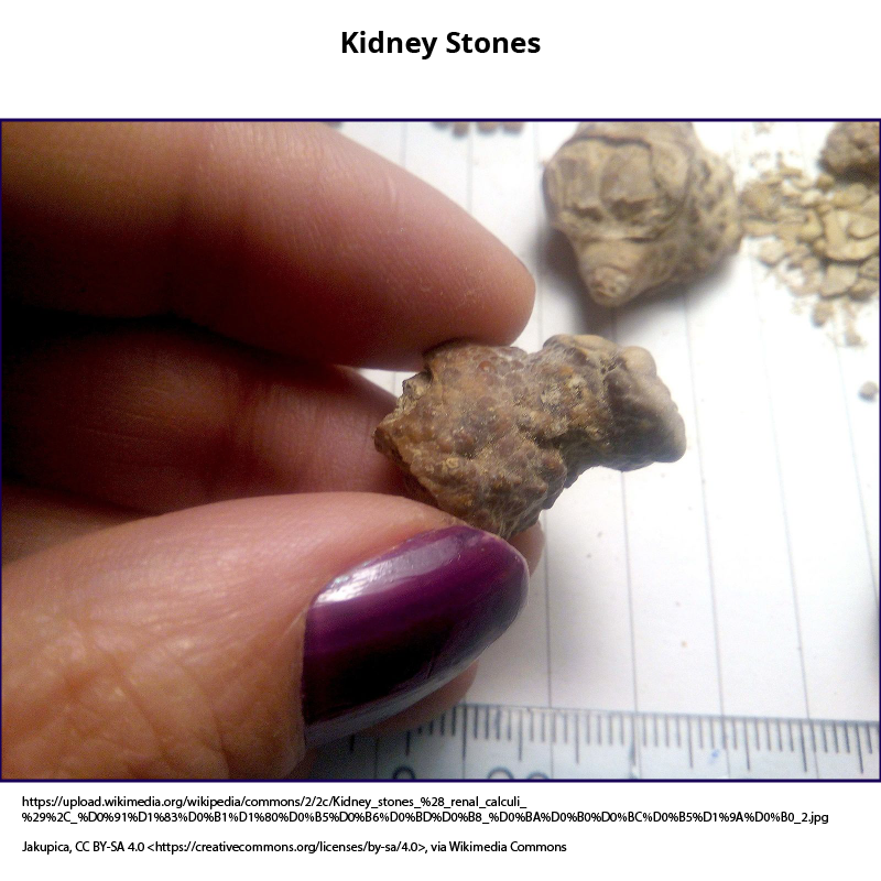
Kidney stones, aka renal calculi, can form within the kidneys or anywhere along the urinary tract; these obstructions can become quite painful and can even lead to permanent damage.
The right and left adrenal, aka suprarenal glands sits on the superior poles of the kidneys.
The hilum is on the medial aspect of the kidney; this is where the renal vessels, nerves, and ureters enter and exit the kidney.
The renal arteries are deeper, and branch directly off the abdominal aorta; notice that the right renal artery travels behind the inferior vena cava.
The renal veins are superficial to the arteries, and drain directly into the inferior vena cava; notice that the left renal vein passes under the superior mesenteric artery.
Innervation is achieved via the renal plexus, which contains sympathetic fibers (from spinal segments T10-12) and parasympathetic fibers (from the vagus nerve).
The ureters descend through the abdominal cavity and into the pelvis; notice that they travel over the iliac vessels before draining urine into the posterolateral walls of the urinary bladder. The ureters are retroperitoneal and are approximately 25 cm long.
The urinary bladder is mostly retroperitoneal (some texts refer to it as subretroperitoneal or infraperitoneal because of its position in the pelvic cavity). It sits immediately posterior to the pubic symphysis of the pelvis; when present, the vagina sites immediately posterior to the urinary bladder and the uterus is superior and posterior.
Urine exits the body via the urethra, which is a thin-walled fibromuscular tube regulated by internal and external sphincters.
The internal sphincter is located in the neck of the bladder at the origin of the urethra; it comprises involuntary smooth muscle and regulates the passage of urine from the urinary bladder to the urethra.
The external sphincter comprises voluntary skeletal muscle fibers derived from the urogenital diaphragm. This is the muscle that we learn to control when we learn to control where and when we void our bladders (toilet training).
Anatomy of micturition (urination).
In females, the urethra is relatively short, 3-5 cm long; it opens to the external environment via the external urethral orifice located in the perineum (not shown in our diagram).
In males, the urethra is longer because it travels the length of the penis; the external urethral orifice is located at the distal end of the penis. The male urethra also provides passage for sperm; we learn more about this in a separate tutorial.
Kidney stones, aka renal calculi, can form within the kidneys or anywhere along the urinary tract; these obstructions can become quite painful and can even lead to permanent damage.
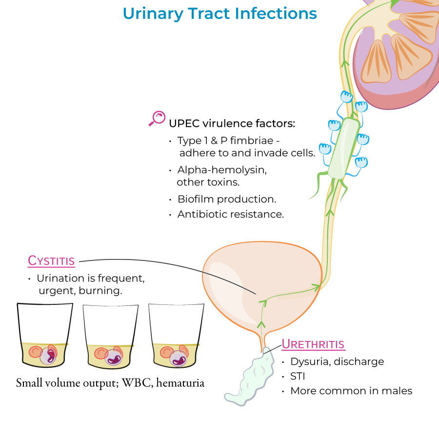
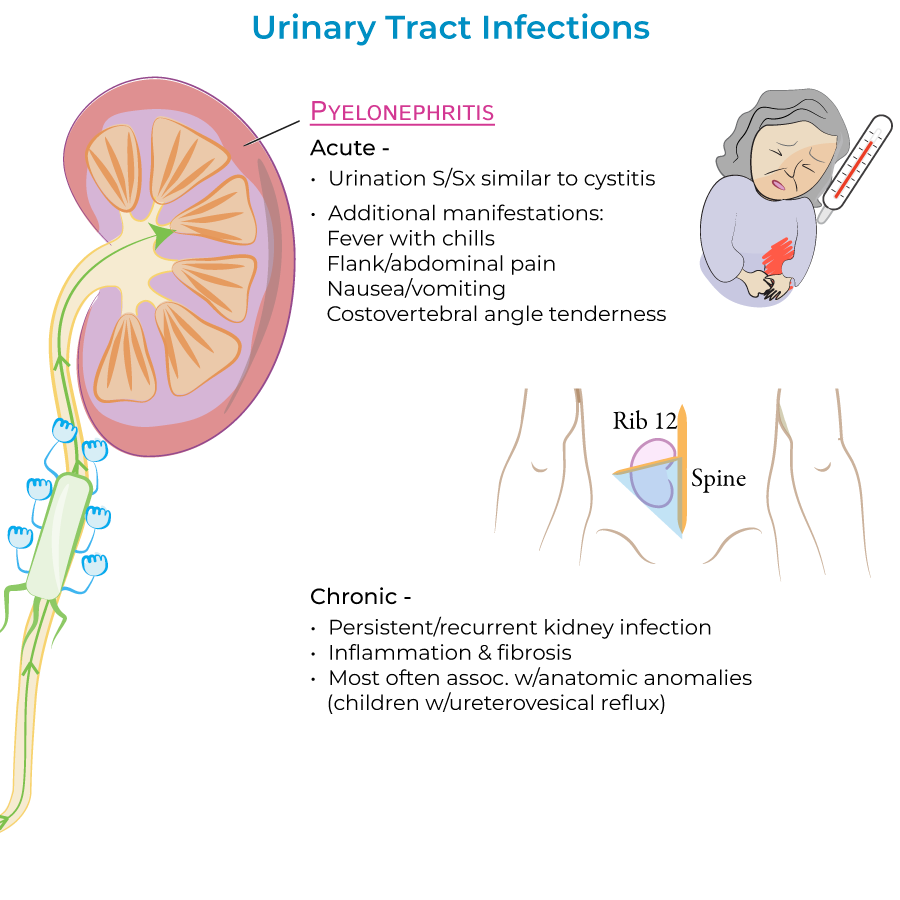
 Urinary tract infections (UTI's) are most commonly caused by the bacteria E. coli; they most often affect the lower urinary tract (the urethra and bladder) but can also ascend to the upper tract (the ureters and kidneys). UTI's are more common in women because their shorter urethras give pathogens quicker access to the urinary bladder.
 Polycystic kidney disease
Urinary incontinence
Urinary System Tumors
Polycystic kidney disease
Urinary incontinence
Urinary System Tumors
Key Structures Overview
Functions
Anatomy
Urinary System
The bean-shaped kidneys, which are retroperitoneal organs in the posterior abdominal wall at approximately level T12-L3.
Notice that the right kidney is slightly lower than the left; this is to accommodate the liver, which lies just beneath the diaphragm on the right side.
The right and left adrenal, aka suprarenal glands sits on the superior poles of the kidneys.
The hilum is on the medial aspect of the kidney; this is where the renal vessels, nerves, and ureters enter and exit the kidney.
The renal arteries are deeper, and branch directly off the abdominal aorta; notice that the right renal artery travels behind the inferior vena cava.
The renal veins are superficial to the arteries, and drain directly into the inferior vena cava; notice that the left renal vein passes under the superior mesenteric artery.
Innervation is achieved via the renal plexus, which contains sympathetic fibers (from spinal segments T10-12) and parasympathetic fibers (from the vagus nerve).
The ureters descend through the abdominal cavity and into the pelvis; notice that they travel over the iliac vessels before draining urine into the posterolateral walls of the urinary bladder. The ureters are retroperitoneal and are approximately 25 cm long.
The urinary bladder is mostly retroperitoneal (some texts refer to it as subretroperitoneal or infraperitoneal because of its position in the pelvic cavity). It sits immediately posterior to the pubic symphysis of the pelvis; when present, the vagina sites immediately posterior to the urinary bladder and the uterus is superior and posterior.
Urine exits the body via the urethra, which is a thin-walled fibromuscular tube regulated by internal and external sphincters.
The internal sphincter is located in the neck of the bladder at the origin of the urethra; it comprises involuntary smooth muscle and regulates the passage of urine from the urinary bladder to the urethra.
The external sphincter comprises voluntary skeletal muscle fibers derived from the urogenital diaphragm. This is the muscle that we learn to control when we learn to control where and when we void our bladders (toilet training).
Anatomy of micturition (urination).
In females, the urethra is relatively short, 3-5 cm long; it opens to the external environment via the external urethral orifice located in the perineum (not shown in our diagram).
In males, the urethra is longer because it travels the length of the penis; the external urethral orifice is located at the distal end of the penis. The male urethra also provides passage for sperm; we learn more about this in a separate tutorial.
Clinical Correlations
Urinary tract infections (UTI's) are most commonly caused by the bacteria E. coli; they most often affect the lower urinary tract (the urethra and bladder) but can also ascend to the upper tract (the ureters and kidneys). UTI's are more common in women because their shorter urethras give pathogens quicker access to the urinary bladder.
Polycystic kidney disease
Urinary incontinence
Urinary System Tumors