The small intestine is a long convoluted segment of the GI tract. It occupies a significant portion of the abdominal cavity, at approximately 20 feet (6-7 meters) in a cadaver, or, due to muscle tone, 10-16 feet (3-5 meters) in a living person.
Functions
The small intestine completes chemical
digestion via digestive juices created by the liver, pancreas, and the small intestine itself.
It absorbs 90% of ingested nutrients and the majority of the water.
The small intestine comprises three continuous segments:
The duodenum, jejunum, and ileum (we can remember the order with the acronym Don’t Jiggle It).
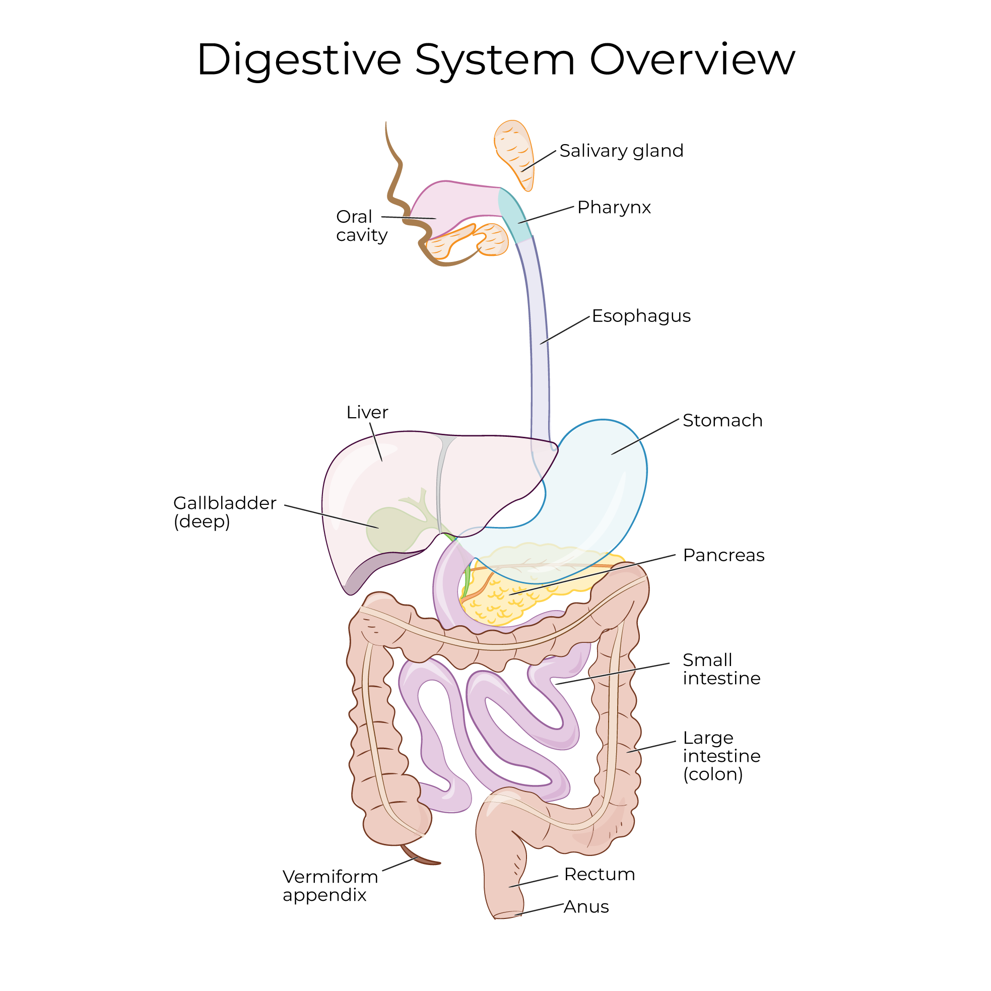
We show some features that are anatomically and physiologically related to the small intestine: the distal end of the stomach, which is continuous with the small intestine, and the pancreas and gallbladder, which send pancreatic juice and bile to the small intestine.
The first segment of the small intestine is the duodenum. It extends superiorly from the pyloric orifice of the stomach and then wraps in a c-shape around the head of the pancreas. It is approximately 10 inches long.
The duodenum is continuous with the jejunum at duodenojejunal flexure. This transition point is attached to the posterior abdominal wall via the suspensory ligament of the duodenum (ligament of Treitz).
In the proximal duodenum, pancreatic juice and bile enter the small intestine via two small openings:
The minor duodenal papilla, which is where the accessory pancreatic duct drains, and the major duodenal papilla, which is where the main pancreatic duct and the bile duct drain.
The passage of bile and pancreatic juices through the major duodenal papilla is regulated by the hepatopancreatic sphincter (aka the Sphincter of Oddi).
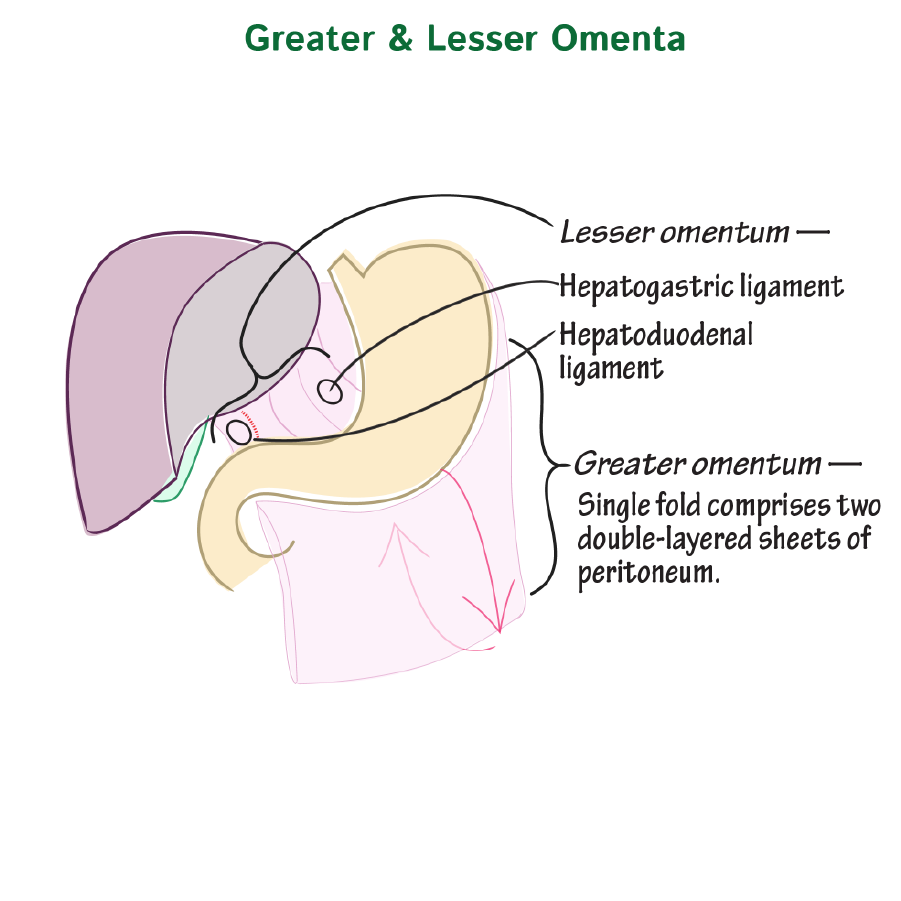
The proximal part of the duodenum is intraperitoneal and connects to the liver via the hepatoduodenal ligament (a part of the lesser omentum), while the remainder is retroperitoneal.
The next two segments are intraperitoneal, and hang suspended from the fan-shaped mesentery proper. The mesentery proper is a fan-shaped, double fold of peritoneum that suspends the intestines from the posterior abdominal wall. Nerves and blood vessels course through this fold to reach the intestines; the mesentery proper is also an important site for fat storage.
The jejunum is the second segment of the small intestine; it is approximately 8 feet long and is the primary site of nutrient absorption.
The ileum is approximately 12 feet long. The ileum terminates at the ileocecal junction, where it is continuous with the cecum of the large intestine; the ileocecal valve regulates passage from the small intestine to the large intestine.
Show that the large intestine wraps around the small intestine in an inverted-U shape.
The greater omentum is a highly vascularized, fatty, apron-like fold of visceral peritoneum that extends from the greater curvature of the stomach and drapes over the small intestine before turning back and ascending to enclose the transverse colon.
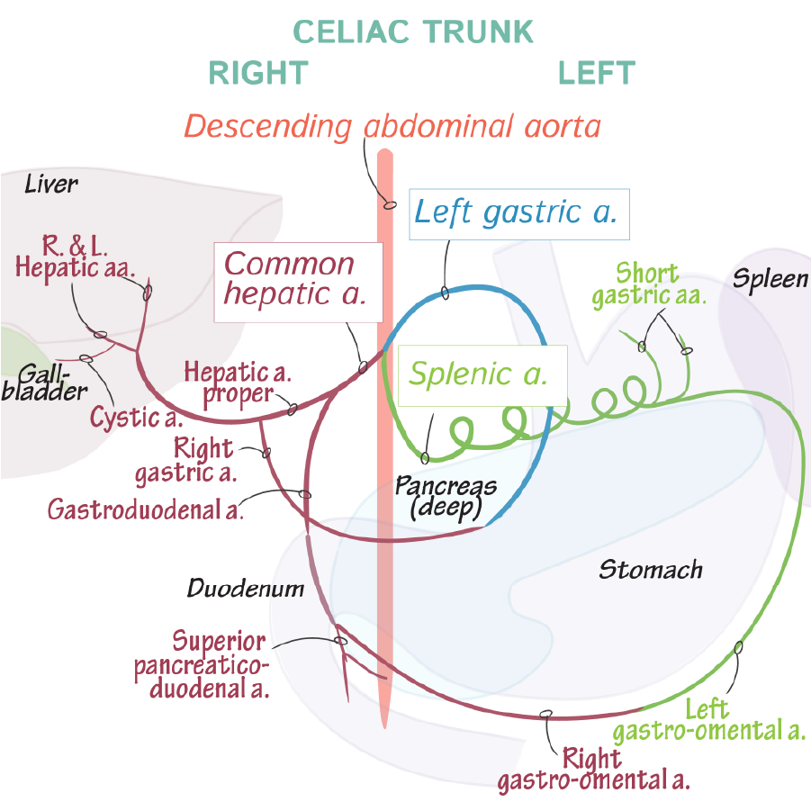
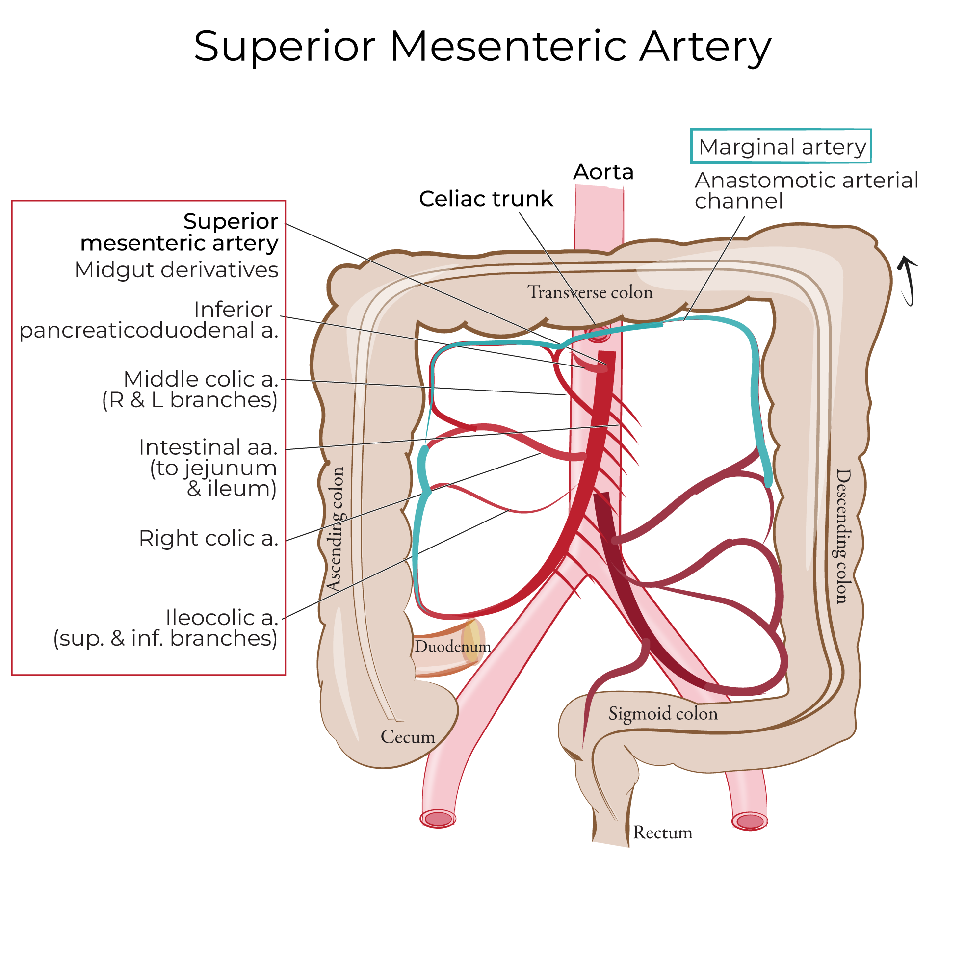
Blood supply to the small intestine is primarily via the superior mesenteric artery, though the celiac artery supplies part of the duodenum.
Innervation is supplied via the superior mesenteric plexus.
To facilitate maximal nutrient and water reabsorption, the small intestine has features that increase its total surface area. The total absorptive surface area of the small intestine is 200-300 sq meters.
The mucosa and submucosa of the small intestine form circular folds that spiral around the diameter of the small intestine, and, at several millimeters tall, are visible with the naked eye. Circular folds increase the surface area of the small intestine and slow down the movements of its contents; they are most prevalent in the distal duodenum and jejunum.
The circular folds have microscopic villi, which are finger-like projections of mucosa.
Intestinal glands reside between the villi; these glands secrete 1-2 liters of intestinal juice daily. Intestinal juice contains mucus, water, electrolytes, and some digestive enzymes.
Each villus is covered by microvilli, which further increase the surface area; collectively, we refer to the microvilli as the intestinal brush border. The brush border produces enzymes that promote digestion.
Review Small Intestine Histology
Meckel’s diverticulum is an embryological remnant of the vitelline duct that manifests as a small pouch in the intestinal wall near where the small and large intestines meet. Meckel’s diverticulum is often asymptomatic, but it can bleeding, inflammation, and obstruction.
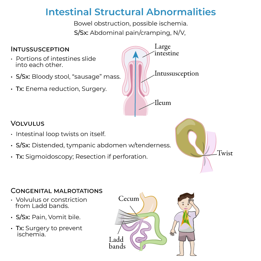
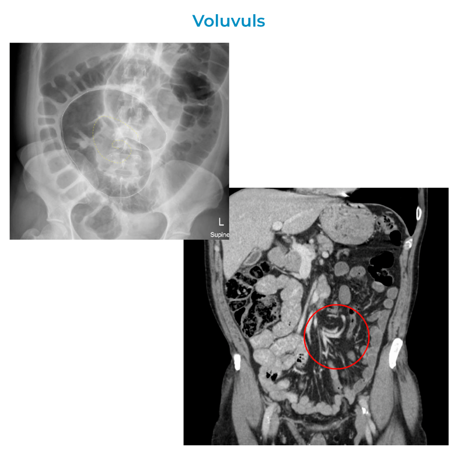
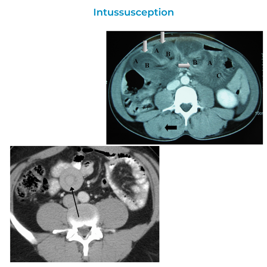
Other
structural abnormalities include intussusception, volvulus, and congenital malrotations.
Additional pathologies