

Start your One-Week Free Trial
Already subscribed? Log in »
Medial Thigh Muscles
Review & Additional Images
Muscles
- Originates as circular muscle from the margin of the obturator foramen and membrane, then forms a tail-like shape as it sends fibers posteriorly behind the femur.
- Inserts on the trochanteric fossa.
- Because it passes anterior to posterior behind the femur, it laterally rotates the thigh.
- Inserts on the medial proximal femur.
- It adducts and flexes the thigh.
- Originates on the inferior ramus and body of the pubis.
- Inserts on the medial surface of the proximal tibia; its tendon merges with the tendons of sartorius and semitendinosus to form the "pes anserinus" ("goose's foot in latin) – the three tendons have a similar appearance to the three-pronged foot of a goose.
- Gracilis adducts the thigh, and, because it crosses the knee, it flexes the leg and medially rotates the leg when the knee is flexed, which is important during gait.

Adductor magnus
- The hamstrings portion originates on the ischial tuberosity and the adductor portion arises from the ischiopubic ramus.
- The hamstrings portion inserts at the adductor tubercle; the adductor portion fans laterally to insert along the posterior femur on the lateral lip of the linea aspera.
- There are small perforations within the muscle near the bone to allow for passage of branches of the deep artery of the thigh.
- The adductor magnus collectively adducts the thigh; the adductor portion flexes the thigh and the hamstrings portion extends it.
- The adductor hiatus is an opening between the adductor portion attachment to the medial supracondylar ridge and the adductor attachment at the adductor tubercle. Indicate the femoral artery and vein as they pass posteriorly to become the popliteal vessels.
 Adductor brevis
- Originates on the pubic body and inferior ramus.
- Inserts on the proximal medial lip of the linea aspera.
- It adducts and flexes the thigh.
 Adductor longus
- Originates on the pubic body.
- Inserts on the linea aspera inferior to adductor brevis.
- It adducts and flexes the thigh.

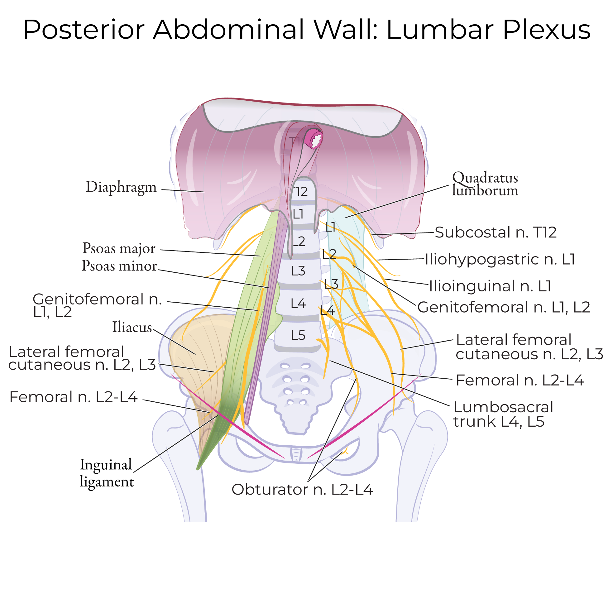
Innervation
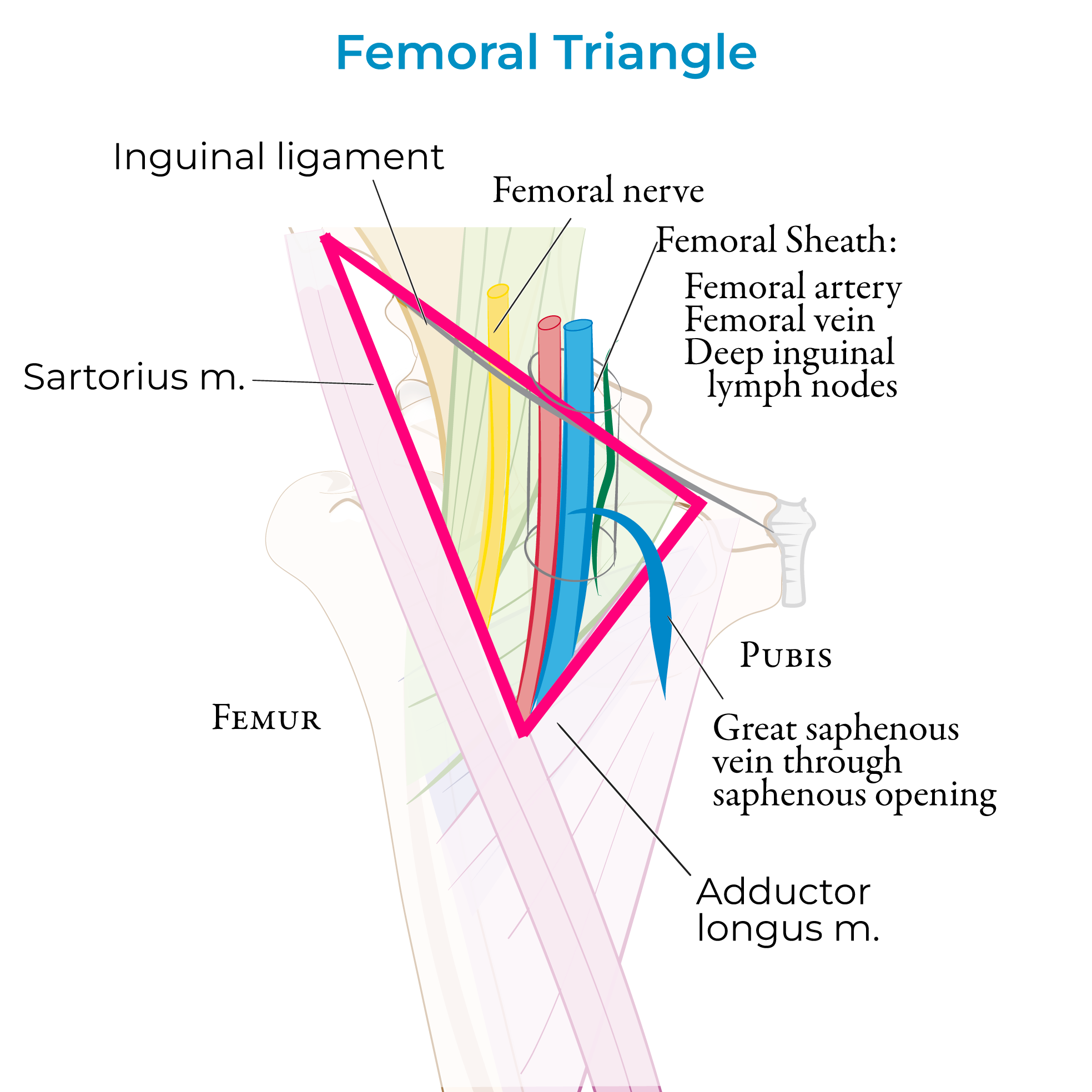
Femoral Triangle
Femoral Triangle Boundaries
The inguinal ligament forms the superior boundary; it forms the base of the triangle.
The lateral border of adductor longus forms the medial border.
Sartorius forms the lateral border of the triangle; the apex is formed by the crossing of sartorius over adductor longus.
The floor of the triangle comprises the muscles iliopsoas and pectineus.
The roof of the triangle is formed by the fascia lata of the thigh (along with the cribriform fascia, subcutaneous tissues, and skin of the thigh). The fascia lata comprises a thick band of connective tissue that envelops the muscles of the thigh.
Contents of the Femoral Triangle
 Neurovascular and lymphatic structures pass through the space deep to the inguinal ligament between the pelvis and the thigh; this space is called the retro-inguinal space.
The vessels bisect the femoral triangle on their way to the adductor canal to traverse the lower thigh.
Mnemonic, from lateral to medial
N - Femoral Nerve and terminal branches.
A - Femoral Artery and its branches.
V - Femoral Vein
E - Empty space (the femoral canal – see notes below)
L - Deep Lymph nodes
Neurovascular and lymphatic structures pass through the space deep to the inguinal ligament between the pelvis and the thigh; this space is called the retro-inguinal space.
The vessels bisect the femoral triangle on their way to the adductor canal to traverse the lower thigh.
Mnemonic, from lateral to medial
N - Femoral Nerve and terminal branches.
A - Femoral Artery and its branches.
V - Femoral Vein
E - Empty space (the femoral canal – see notes below)
L - Deep Lymph nodes
Femoral Sheath:
The femoral sheath is a 3-4 cm long fascial tube derived from transversalis and iliac fascia. The sheath envelops the femoral artery and vein and their branches, as well as the deep inguinal lymph nodes.
The fascial sheath itself is divided into compartments:
Lateral, containing the femoral artery.
Intermediate, containing the femoral vein.
Medial, which houses the femoral canal with lymph vessels.
Great Saphenous Vein & Clinical Correlations
The great saphenous vein, which is the long superficial vein that drains the lower extremity, passes through the saphenous opening of the fascia lata to drain into the femoral vein within the femoral triangle.
The great saphenous vein is often used in arterial bypass operations (coronary artery and peripheral), due to its large size and relatively easy access.
In an emergency setting, the great saphenous vein can be used for venous cutdown, in which the vein is exposed and a cannula or catheter is inserted into the vessel.