

Start your One-Week Free Trial
Already subscribed? Log in »

Forearm - Posterior Muscles
Here we'll learn the muscles of the posterior forearm, which extend and adduct the wrist and digits (with a couple of notable exceptions). These muscles are innervated by the radial nerve and its branches.
We'll start with the superficial layer of muscles, which are named after their locations and their actions: extensor carpi radialis longus, extensor carpi radialis brevis, extensor digitorum, extensor digiti minimi, and extensor carpi ulnaris.
A lateral band of connective tissue called the extensor retinaculum passes over the tendons of the posterior forearm at the wrist and holds them in place.
A triangular shaped depression in the skin at the base of the thumb.
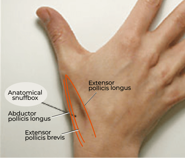 The lateral and medial borders of the "snuffbox" are created by the tendons of abductor pollicis longus, extensor pollicis longus, and extensor pollicis brevis; its floor is bound by the scaphoid and trapezium bones.
Key structures passing through the anatomical snuffbox can be remembered with the mnemonic CARTS:
The Cephalic vein (which forms from the dorsal venous network of the hand)
The radial Artery (a branch of the brachial artery)
The Radial nerve, Superficial branch,
And, the Tendons of extensor carpi radialis longus and brevis.
Because the superficial branch of the radial artery passes through here, a pulse can be felt in the anatomical snuffbox.
And, because the scaphoid bone forms the floor of the snuffbox, fractures to this carpal bone produce pain and tenderness to the area.
The lateral and medial borders of the "snuffbox" are created by the tendons of abductor pollicis longus, extensor pollicis longus, and extensor pollicis brevis; its floor is bound by the scaphoid and trapezium bones.
Key structures passing through the anatomical snuffbox can be remembered with the mnemonic CARTS:
The Cephalic vein (which forms from the dorsal venous network of the hand)
The radial Artery (a branch of the brachial artery)
The Radial nerve, Superficial branch,
And, the Tendons of extensor carpi radialis longus and brevis.
Because the superficial branch of the radial artery passes through here, a pulse can be felt in the anatomical snuffbox.
And, because the scaphoid bone forms the floor of the snuffbox, fractures to this carpal bone produce pain and tenderness to the area.
Superficial Layer
Extensor carpi radialis longus
- Arises from the supracondylar ridge of the humerus and inserts on the base of metacarpal II.
- Extensor carpi radialis longus extends and abducts the wrist.
Common extensor tendon injury
Overuse injury to this tendon, lateral epicondylitis, is referred to as "tennis elbow" because it is common in those who play tennis or engage in other activities with repetitive wrist extension.
Extensor carpi radialis brevis
- Inserts on the base of metacarpal III.
- Has the same actions, wrist extension and abduction, as extensor carpi radialis longus.
Extensor digitorum
- Gives rise to four tendons that split to form the extensor central and lateral bands, which insert on digits 2-5.
- The extensor central bands insert on the middle phalanges, and the extensor lateral bands insert on the distal phalanges; the bands are wrapped in the connective tissues of the extensor hoods (aka extensor expansions).
- Extensor digitorum, as its name suggests, extends the wrist and digits 2-5.
Extensor digiti minimi
- Inserts into the extensor hood of digit 5, the little, aka, minimi, finger, which it extends.
Extensor carpi ulnaris
- Arises from the common extensor tendon and the posterior border of the ulna, and inserts on the base of metacarpal 5.
- Extensor carpi ulnaris extends the wrist.
- Because it inserts medially, it adducts the wrist.
Deep Layer
Supinator,
- This is a broad, flat muscle with two heads.
- The superficial head arises from the lateral epicondyle of the humerus and associated ligaments, and the deeper head arises from the supinator crest of the ulna.
- The two heads wrap around the proximal radius to insert on the anterior, lateral, and proximal aspects of the upper 1/3rd of the radius.
- This spiral formation provides forearm supination, which brings the palms of the hands upwards (remember: "hold the SOUP" for supination; "POURing the soup for pronation").
Abductor pollicis longus
- Arises from the middle third of the posterior radius and ulna and inserts via on the base of metacarpal 1.
- Because it crosses the wrist from medial to lateral, this muscle abducts digit 1.
Extensor pollicis brevis
- Arises from the radius and interosseous membrane and inserts on the base of the proximal phalanx of digit 1, which it extends.
Extensor pollicis longus
- Arises from the ulna and interosseous membrane and inserts on the base of the distal phalanx of digit 1, which it extends.
Extensor indicis
- Arises from the ulna and interosseous membrane and inserts via a long tendon to the extensor hood of digit 2, the "pointer finger," which it extends.
Extensor retinaculum
Anatomical Snuffbox
The lateral and medial borders of the "snuffbox" are created by the tendons of abductor pollicis longus, extensor pollicis longus, and extensor pollicis brevis; its floor is bound by the scaphoid and trapezium bones.
Key structures passing through the anatomical snuffbox can be remembered with the mnemonic CARTS:
The Cephalic vein (which forms from the dorsal venous network of the hand)
The radial Artery (a branch of the brachial artery)
The Radial nerve, Superficial branch,
And, the Tendons of extensor carpi radialis longus and brevis.
Because the superficial branch of the radial artery passes through here, a pulse can be felt in the anatomical snuffbox.
And, because the scaphoid bone forms the floor of the snuffbox, fractures to this carpal bone produce pain and tenderness to the area.