Here we’ll learn about the arteries of the upper extremity.
To begin, we draw the relevant musculoskeletal components; we focus on the right side of the body.
Show two muscular landmarks:
Pectoralis minor extends from the anterior surfaces of ribs 3-5 to the coracoid process of the scapula.
Teres major arises from the inferior angle of the scapula and inserts on the medial lip of the intertubercular sulcus of the humerus.
Now we’re ready to show the vessels.
For context, start with the heart and show the aorta arching up and then descending through the thoracic cavity.
Branches of the arch of the
aorta:
The brachiocephalic trunk gives rise to the right common carotid and subclavian arteries.
The left common carotid and left subclavian arteries then branch separately from the arch.
Now we’ll follow the course of the right subclavian artery as it gives rise to the arteries of the upper extremity; this is mirrored on the left.
The subclavian artery becomes the axillary artery at the lateral margin of rib 1.
The axillary artery gives off 6 branches and can be divided into three parts depending on its relation to the pectoralis minor muscle.
Proximal to pectoralis minor: the superior thoracic artery (this vessel is variable and may anastomose with the intercostal and internal thoracic arteries).
Two vessels arise from under pectoralis minor:
The thoraco-acromial artery is a short trunk that pierces the costocoracoid membrane and gives rise to 4 branches (acromial, deltoid, pectoral, clavicular).
The lateral thoracic artery begins near the inferior edge of pectoralis; this vessel supplies chest muscles and the lateral breast (note that the lateral thoracic artery may arise from thoraco-acromial artery).
Lastly, in the section distal to pectoralis minor, the subscapular artery and the anterior and posterior circumflex humeral arteries.
The subscapular artery is short and wide; it descends along the lateral edge of subscapularis and divides into circumflex scapular and thoracodorsal arteries.
The circumflex humeral arteries encircle the surgical neck of the humerus to anastomose laterally; they may arise as separate vessels or together as a common trunk that splits.
The anterior branch is smaller and passes deep to coracobrachialis and biceps brachii; the posterior branch is larger and passes through the quadrangular space with the axillary nerve. These vessels supply the glenohumeral joint and nearby muscles.
The axillary artery becomes brachial artery at the inferior margin of teres major; the brachial artery travels in the anterior compartment to the cubital fossa.
Because it is easily accessible and close to the heart, the brachial artery is often used to measure
blood pressure.
The largest branch of the brachial artery is the profunda brachii artery (aka deep brachial artery); it passes through the triangular interval (with the radial nerve) to serve the posterior compartment of the arm.
On the posterior surface of the humerus, the profunda brachii artery travels within the radial groove; it terminates by splitting to form the radial and middle collateral arteries, which anastomose with vessels around the elbow (we’ll see these soon).
The brachial artery also gives off two medial branches that travel to the elbow; these are the ulnar collateral arteries.
The brachial artery terminates in the inferior cubital fossa, where it splits to form the radial and ulnar arteries.
The radial artery travels along the lateral side of the forearm; proximally it is deep to brachioradialis, but distally it is palpable under the skin between the tendons of brachioradialis and flexor carpi radialis. You can read about the radial arterial pulse and Allen’s test in our notes.
In the wrist, the radial artery passes over the floor of the anatomical snuffbox to enter the hand, where it gives rise to the deep palmar arch and several other branches that serve the wrist and hand.
The radial artery sends a branch, called the recurrent radial artery, to anastomose with the radial collateral artery at the elbow.
The radial artery supplies the muscles of the lateral sides of the anterior forearm, wrist, and hand.
Now, show that the ulnar artery runs along the medial side of the forearm; it passes between the deep and intermediate muscle layers.
At the wrist, the ulnar artery passes superficial to the flexor retinaculum and travels with the ulnar nerve in the ulnar canal (Guyon’s canal) to enter the hand where it gives rise to the superficial palmar arch.
The ulnar artery has several branches that supply the elbow and forearm:
Two ulnar recurrent arteries travel superiorly to anastomose with the ulnar collateral arteries.
Iterosseous arteries branch from a common stem; they provide blood to the anterior and posterior compartments of the forearm.
The ulnar artery primarily supplies the medial (ulnar) side of the hand, including the little finger and medial half of the ring finger.
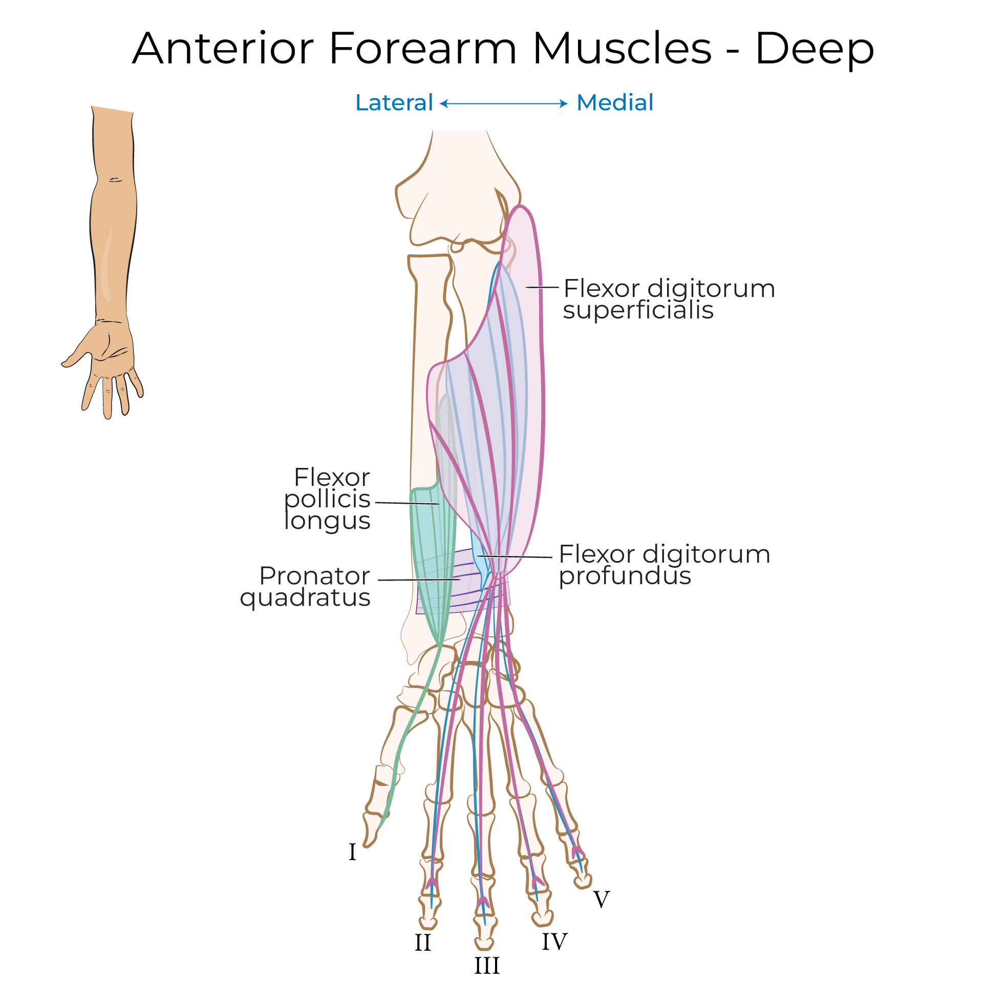
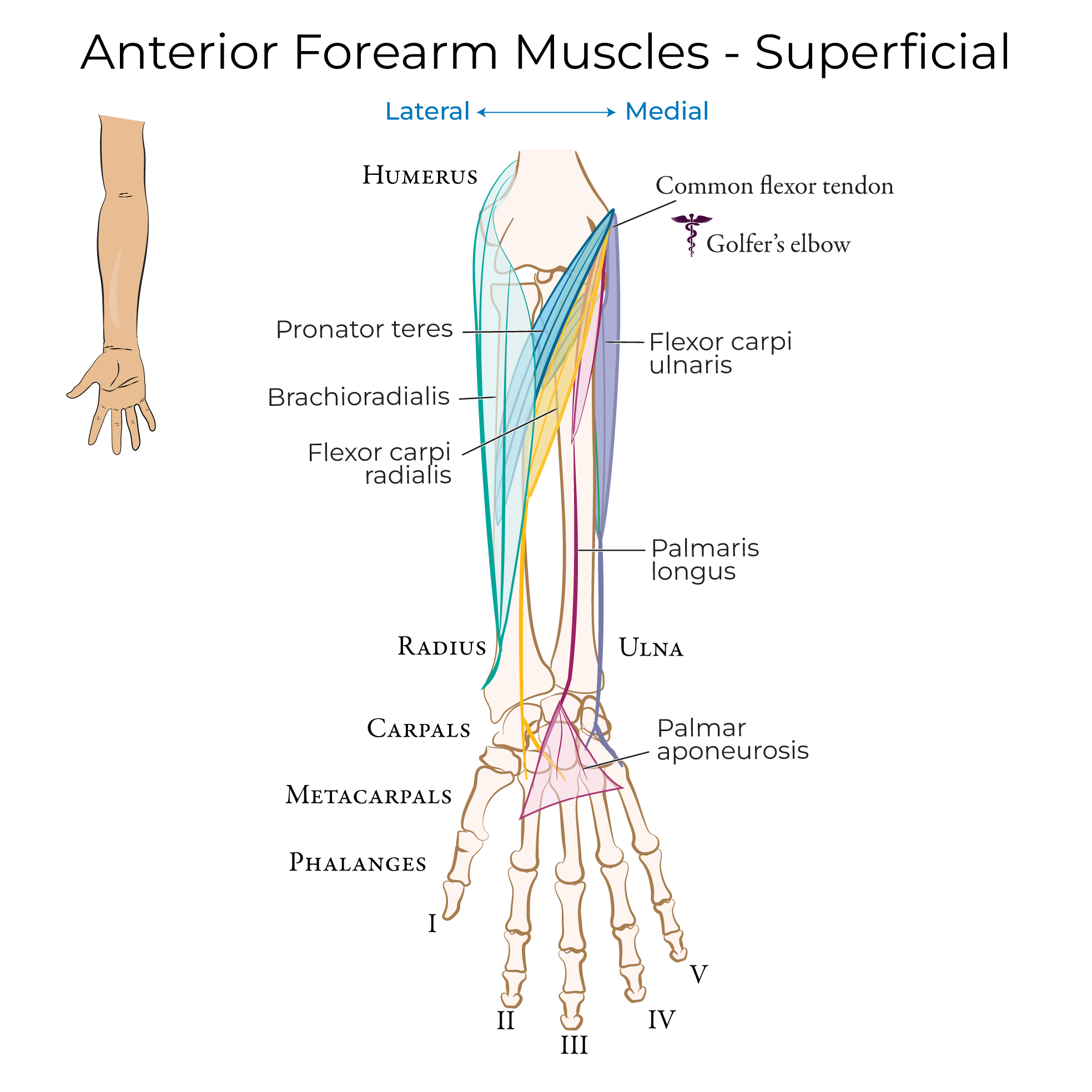
Now, let’s draw a more detailed diagram of the forearm to show key relationships between the arteries and muscles.
The profunda brachii artery splits to give rise to the radial collateral and middle collateral arteries.
Medially, show that the brachial artery gives off the superior and inferior ulnar collateral arteries.
The brachial artery gives rise to the radial and ulnar arteries after it passes through the cubital fossa.
Now we can show the anastomoses around the elbow.
The recurrent radial artery travels proximally and anastomoses with the radial collateral artery.
On the ulnar side, the anterior ulnar recurrent artery travels proximally and anastomoses with the inferior ulnar collateral artery.
The posterior ulnar recurrent artery travels posterior to the humeral epicondyle and anastomoses with the superior ulnar collateral artery.
A helpful mnemonic for remembering these anastomoses is, “ I Am So Perceptive”:
The Inferior ulnar collateral artery meets the Anterior ulnar recurrent artery;
The Superior ulnar collateral artery meets the Posterior ulnar recurrent artery.
The ulnar artery gives rise to the interosseous arteries as follows:
The common interosseous artery is short, and quickly divides to form the anterior and posterior interosseous arteries.
The anterior interosseous artery travels distally along the interosseous membrane (with the anterior interosseous nerve). When it reaches the proximal border of pronator quadratus, it pierces the interosseous membrane and moves posteriorly.
The posterior interosseous artery travels between the deep and superficial extensor muscles (with the posterior interosseous nerve). Before diving posteriorly, the posterior interosseous artery gives off the recurrent interosseous artery, which travels proximally and passes posteriorly to anastomose with the middle collateral artery.
The ulnar artery enters the hand and forms the superficial palmar arch, which is superficial to the tendons of flexor digitorum profundus.
The radial artery enters the hand and forms the deep palmar arch deep to the tendons.
Within the wrist and hand, the radial and ulnar arteries give rise to several smaller branches and anastomoses that serve the palm and digits.


