

Start your One-Week Free Trial
Already subscribed? Log in »
Internal Iliac artery branches
Here we will learn the branches of the internal iliac artery, which carry oxygenated blood to the pelvic viscera and musculoskeletal structures, the gluteal and medial thigh regions, and the perineum.
Be aware that these branching patterns are variable, and some are contested.
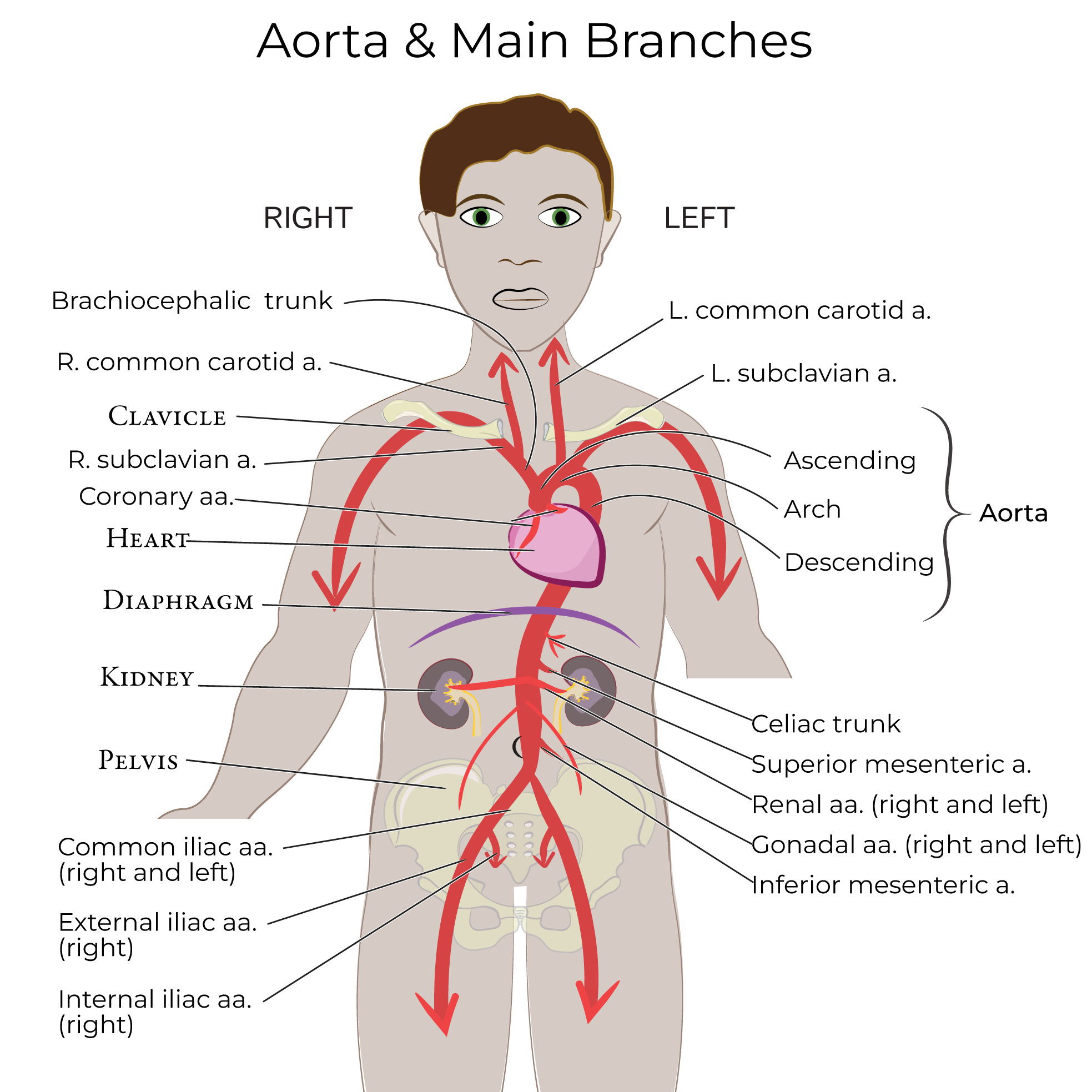
Origins of the internal iliac artery: the right common iliac artery descends along the anterior surface of the sacrum and divides to form the external iliac artery, which exits anteriorly to supply the lower extremity, and, the internal iliac artery, which enters the pelvis.
 The internal iliac artery splits into anterior and posterior divisions, aka trunks.
 Branches of the anterior division supply the pelvic organs, the perineum, and muscles of the gluteal and adductor regions of the lower extremity.
The umbilical artery travels anteriorly; during the fetal period, this vessel traveled to the placenta. Postnatally, remnants of this vessel persist as the medial umbilical ligaments (they travel within the medial umbilical folds in the peritoneum, along the posterior aspect of the abdominal muscles).
The patent part of the umbilical artery gives off a branch, called the superior vesicle artery, which travels to the urinary bladder (when you hear “vesicle” think “urinary bladder”). Note that this artery may arise directly from the internal iliac artery; it may also give rise to a branch that travels to the ductus deferens.
The obturator artery travels along the lateral wall of the pelvis, over the fascia that covers the obturator internus muscle, and exits the pelvis via the obturator canal with the obturator nerve to supply the adductors of the thigh.
The inferior vesicle artery descends to supply the urinary bladder and adjacent reproductive structures (such as the prostate). We note that the vaginal and uterine arteries are also in this area.
The vessels that supply the urinary bladder and reproductive structures are highly variable. In about half of women, the inferior vesicle artery is absent; alternatively, the inferior vesicle artery can be a branch of the vaginal artery. It’s important to be aware of these variations during surgery.
The middle rectal artery anastomoses with the superior and inferior rectal arteries, which branch from the inferior mesenteric and internal pudendal arteries, respectively.
The internal pudendal artery exits the pelvis via the greater sciatic foramen, inferior to piriformis. The internal pudendal artery will enter the perineum via the lesser sciatic foramen; it travels with the pudendal vein and nerve in the pudendal canal.
The inferior gluteal artery is the terminal branch of the anterior division; it exits the pelvis via the sciatic foramen to supply the gluteal region of the posterior hip. Be aware that the inferior gluteal artery may instead originate from the posterior division.
Branches of the posterior division supply the posterior abdominal and pelvic walls and the gluteal region.
The iliolumbar artery travels superiorly and ultimately splits to form iliac and lumbar branches. Alternatively, the iliolumbar artery may arise directly from the internal iliac artery (prior to the anterior/posterior split).
The lateral sacral artery travels along the anterior surface of the sacrum. The lateral sacral artery gives rise to spinal branches that travel through the anterior sacral foramina; it is often paired.
The superior gluteal artery, which exits the pelvic cavity superior to piriformis, via the greater sciatic foramen, to reach the posterior hip. This is a large vessel that supplies the gluteal and pelvic wall muscles.
Branches of the anterior division supply the pelvic organs, the perineum, and muscles of the gluteal and adductor regions of the lower extremity.
The umbilical artery travels anteriorly; during the fetal period, this vessel traveled to the placenta. Postnatally, remnants of this vessel persist as the medial umbilical ligaments (they travel within the medial umbilical folds in the peritoneum, along the posterior aspect of the abdominal muscles).
The patent part of the umbilical artery gives off a branch, called the superior vesicle artery, which travels to the urinary bladder (when you hear “vesicle” think “urinary bladder”). Note that this artery may arise directly from the internal iliac artery; it may also give rise to a branch that travels to the ductus deferens.
The obturator artery travels along the lateral wall of the pelvis, over the fascia that covers the obturator internus muscle, and exits the pelvis via the obturator canal with the obturator nerve to supply the adductors of the thigh.
The inferior vesicle artery descends to supply the urinary bladder and adjacent reproductive structures (such as the prostate). We note that the vaginal and uterine arteries are also in this area.
The vessels that supply the urinary bladder and reproductive structures are highly variable. In about half of women, the inferior vesicle artery is absent; alternatively, the inferior vesicle artery can be a branch of the vaginal artery. It’s important to be aware of these variations during surgery.
The middle rectal artery anastomoses with the superior and inferior rectal arteries, which branch from the inferior mesenteric and internal pudendal arteries, respectively.
The internal pudendal artery exits the pelvis via the greater sciatic foramen, inferior to piriformis. The internal pudendal artery will enter the perineum via the lesser sciatic foramen; it travels with the pudendal vein and nerve in the pudendal canal.
The inferior gluteal artery is the terminal branch of the anterior division; it exits the pelvis via the sciatic foramen to supply the gluteal region of the posterior hip. Be aware that the inferior gluteal artery may instead originate from the posterior division.
Branches of the posterior division supply the posterior abdominal and pelvic walls and the gluteal region.
The iliolumbar artery travels superiorly and ultimately splits to form iliac and lumbar branches. Alternatively, the iliolumbar artery may arise directly from the internal iliac artery (prior to the anterior/posterior split).
The lateral sacral artery travels along the anterior surface of the sacrum. The lateral sacral artery gives rise to spinal branches that travel through the anterior sacral foramina; it is often paired.
The superior gluteal artery, which exits the pelvic cavity superior to piriformis, via the greater sciatic foramen, to reach the posterior hip. This is a large vessel that supplies the gluteal and pelvic wall muscles.
Internal Iliac Artery
The internal iliac artery splits into anterior and posterior divisions, aka trunks.
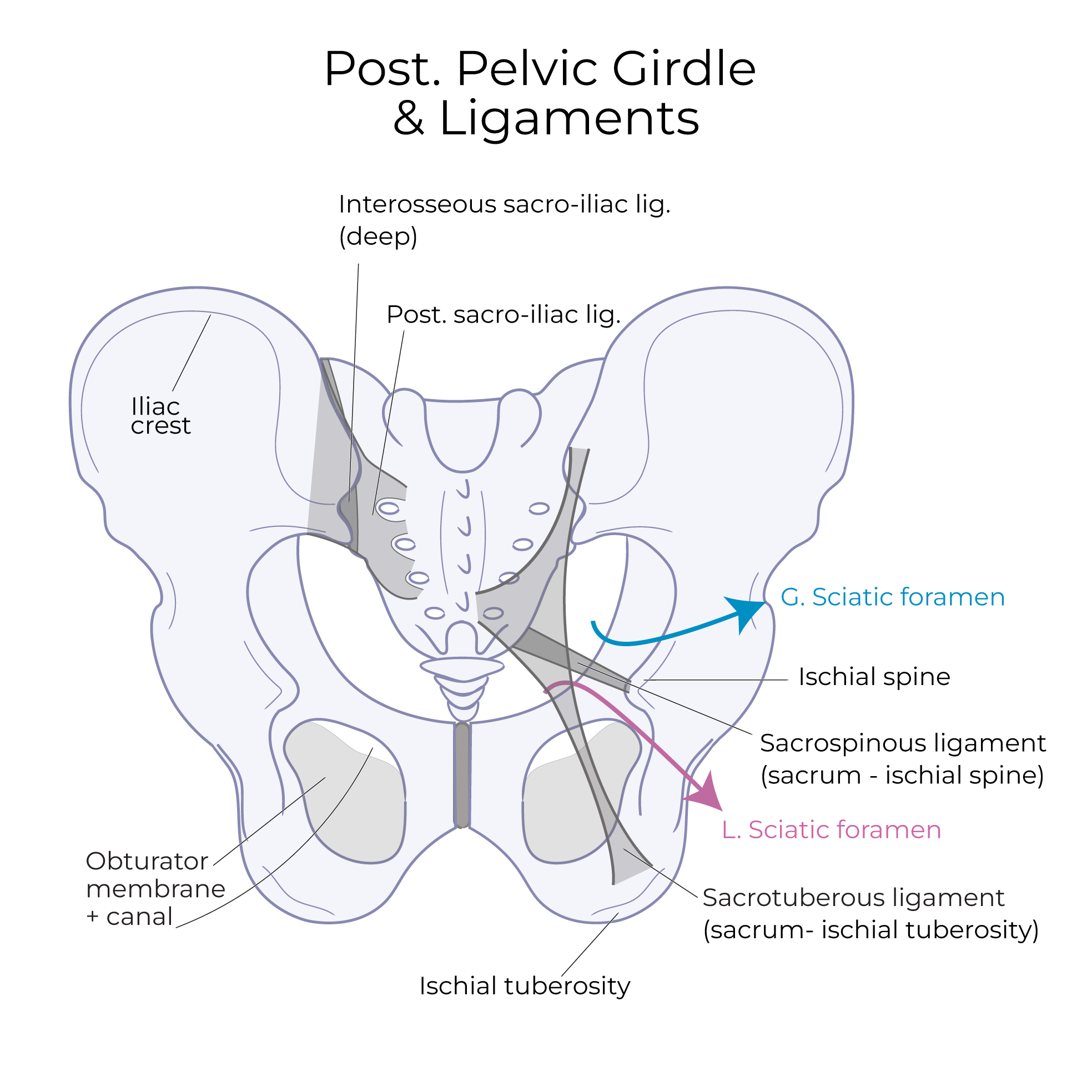
Pelvic girdle:
Draw the os coxa and sacrum, and show the sacrotuberous and sacrospinous ligaments, and the piriformis and obturator internus muscles. Notice we’ve left a small opening, the obturator canal, in the border of obturator internus.
Internal Iliac Artery: Anterior Division
Internal Iliac Artery: Posterior Division