

Start your One-Week Free Trial
Already subscribed? Log in »


Respiratory System Development
Here we’ll learn about the development of the respiratory system from approximately week 4 onward; be aware that timing varies and the stages may overlap. (We’re using the timeline provided by Schoenwolf et al 2023).
Key embryological tissues include:
Endoderm, which gives rise to the epithelial cells of the lower respiratory tract (the lower larynx, trachea, bronchial tree, and alveoli).
Splanchnic mesoderm gives rise to the connective and smooth muscle tissue of the lower respiratory tract, and to the visceral pleura that covers the lungs.
The respiratory system is derived from the ventral wall of the primitive gut tube at approximately week 4, when an outpocket of the foregut forms; this is called the lung bud, aka, respiratory diverticulum.
Week four is the start of the embryological stage of respiratory system development.
During this stage, the respiratory diverticulum forms and gives rise to the laryngeal epithelium (below the vocal folds), the tracheobronchial tree, and lungs.
The respiratory diverticulum forms as an anterior outcropping of the foregut; the tracheoesophageal ridges continue to connect the two structures.
Splanchnic mesoderm surrounds the respiratory diverticulum.
The tracheoesophageal ridges form a septum that separate the primitive foregut into the ventral laryngotracheal tube and dorsal esophagus. We can see the trachea and the primary bronchial buds.
 The pseudoglandular stage begins in approximately week 6 or earlier; this stage is characterized by continuous branching that gives rise to bronchioles, and ultimately, and terminal bronchioles. Branching is regulated via epithelial-mesenchyme interactions and is under the direction of fibroblast growth factor 10 and other signaling molecules.
The terminal bronchioles arise as thick-walled tubes of cuboidal epithelium from the ends of the bronchioles.
In the surrounding mesenchyme, fibroblasts are busy producing components of the surrounding connective tissue, and we can start to see capillaries; these features give the growing tissue a glandular appearance, which is why this is called the pseudoglandular stage.
The conducting portion of bronchial tree is formed by approximately week 16, when the canalicular stage begins.
The terminal bronchioles divide and form multiple respiratory bronchioles, which give rise to alveolar ducts; the walls of the tubules grow thinner as their lumens enlarge. The mesenchyme surrounding the bronchioles becomes increasingly vascularized during this stage. Limited, inefficient gas exchange is possible.
By approximately week 24, we enter the saccular stage of respiratory development.
The walls of the respiratory bronchioles continue to thin as their distal ends widen to form terminal sacs (precursors to alveoli). In the surrounding tissue, we start to see elastic fibers and thin bands of smooth muscle fibers forming. These are crucial for normal lung functioning.
The epithelial lining of the sacs comprises type I and type II cells.
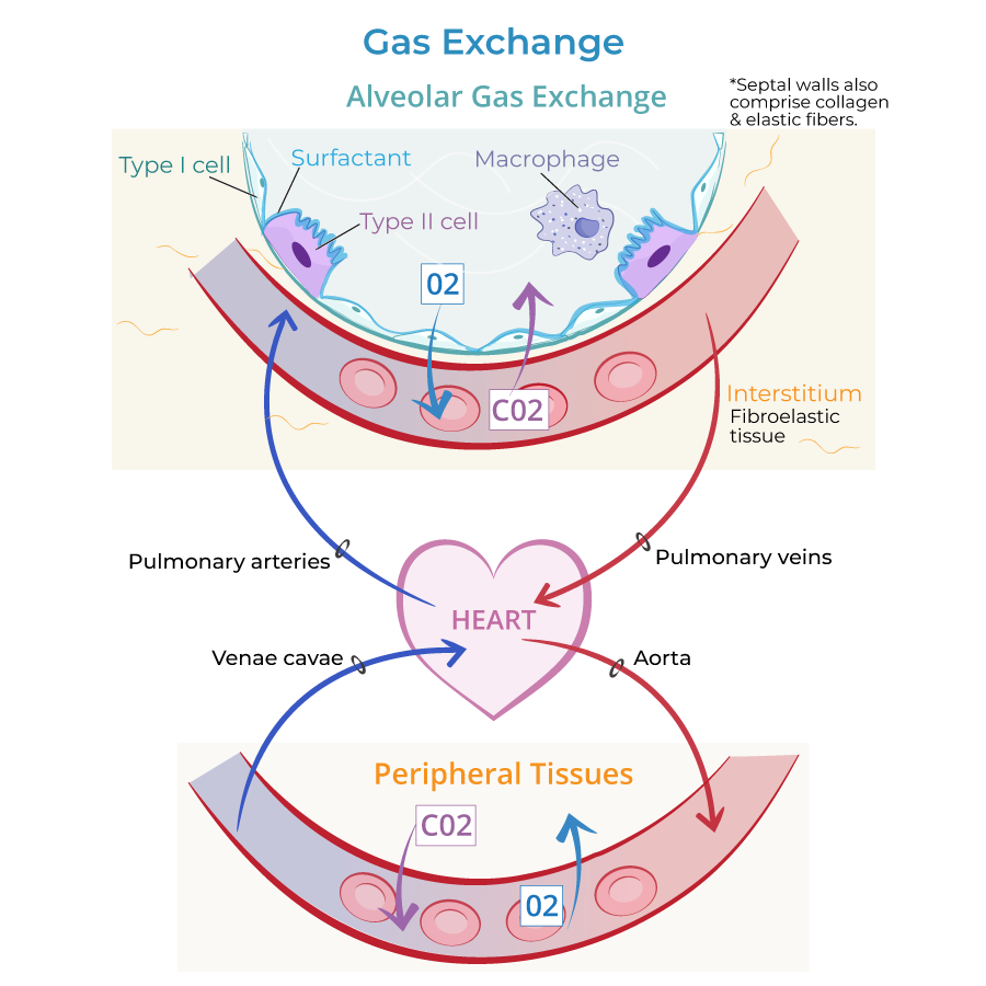
Type I alveolar cells are simple squamous epithelial cells that provide a surface for gas exchange; they cover 90-95% of the alveolar epithelial surface area.
Type II alveolar cells produce pulmonary surfactant, which contains phospholipids and surfactant proteins to prevent alveolar collapse (atelectasis), especially during expiration.
By approximately week 36, the fetus enters the alveolar stage, which is the final stage of respiratory system development.
By approximately 36 weeks, the formation and multiplication of mature alveoli begins; mature alveoli are capable of efficient gas exchange. After birth, septation continues to subdivide the alveoli. This stage lasts throughout childhood, possibly even into early adulthood.
Alveoli arise from the respiratory bronchiole as individual alveoli and as clusters of alveolar sacs.
Along the bronchioles, show pink bands of smooth muscle fibers, and, along the bronchioles and in the walls of the alveoli, show thin green elastic fibers.
Capillary networks surround the alveoli, which allows for efficient gas exchange.
At birth, the lungs comprise 20-50 million primitive alveoli; by adulthood, they comprise 300-500 million mature alveoli.
The pseudoglandular stage begins in approximately week 6 or earlier; this stage is characterized by continuous branching that gives rise to bronchioles, and ultimately, and terminal bronchioles. Branching is regulated via epithelial-mesenchyme interactions and is under the direction of fibroblast growth factor 10 and other signaling molecules.
The terminal bronchioles arise as thick-walled tubes of cuboidal epithelium from the ends of the bronchioles.
In the surrounding mesenchyme, fibroblasts are busy producing components of the surrounding connective tissue, and we can start to see capillaries; these features give the growing tissue a glandular appearance, which is why this is called the pseudoglandular stage.
The conducting portion of bronchial tree is formed by approximately week 16, when the canalicular stage begins.
The terminal bronchioles divide and form multiple respiratory bronchioles, which give rise to alveolar ducts; the walls of the tubules grow thinner as their lumens enlarge. The mesenchyme surrounding the bronchioles becomes increasingly vascularized during this stage. Limited, inefficient gas exchange is possible.
By approximately week 24, we enter the saccular stage of respiratory development.
The walls of the respiratory bronchioles continue to thin as their distal ends widen to form terminal sacs (precursors to alveoli). In the surrounding tissue, we start to see elastic fibers and thin bands of smooth muscle fibers forming. These are crucial for normal lung functioning.
The epithelial lining of the sacs comprises type I and type II cells.
Type I alveolar cells are simple squamous epithelial cells that provide a surface for gas exchange; they cover 90-95% of the alveolar epithelial surface area.
Type II alveolar cells produce pulmonary surfactant, which contains phospholipids and surfactant proteins to prevent alveolar collapse (atelectasis), especially during expiration.
By approximately week 36, the fetus enters the alveolar stage, which is the final stage of respiratory system development.
By approximately 36 weeks, the formation and multiplication of mature alveoli begins; mature alveoli are capable of efficient gas exchange. After birth, septation continues to subdivide the alveoli. This stage lasts throughout childhood, possibly even into early adulthood.
Alveoli arise from the respiratory bronchiole as individual alveoli and as clusters of alveolar sacs.
Along the bronchioles, show pink bands of smooth muscle fibers, and, along the bronchioles and in the walls of the alveoli, show thin green elastic fibers.
Capillary networks surround the alveoli, which allows for efficient gas exchange.
At birth, the lungs comprise 20-50 million primitive alveoli; by adulthood, they comprise 300-500 million mature alveoli.
 Features of Adult Tracheobronchial Tree
Photos of adult respiratory structures
Histology of Respiratory System
Features of Adult Tracheobronchial Tree
Photos of adult respiratory structures
Histology of Respiratory System
Overview
Embryological Stage
Esophageal atresia and tracheoesophageal fistula
Abnormal separation of the foregut into esophagus and trachea; they often co-occur.
By the end of the embryonic stage, the stem of the diverticulum gives rise to the trachea and primary bronchi; further bronchial divisions give rise to the secondary and tertiary bronchi.
With the development of the secondary bronchi, we can start to identify lobes of the lungs: three on the right (superior, middle, and inferior) and two on the left (superior and inferior). We indicate the visceral pleura, which is the covering of the lungs.
The formation of the tertiary bronchi corresponds to the development of bronchopulmonary segments.
Recall that the left lung is smaller to make space for the heart within the thorax.
On a related note, partitions start to form during the fourth week that will ultimately subdivide the body into pericardial, pleural, and peritoneal cavities.
Pseudoglandular Stage
Canalicular Stage
Saccular Stage
RDS
Surfactant deficiency can lead to neonatal respiratory distress syndrome (RDS), which is characterized by progressive respiratory failure with alveolar collapse, reduced lung compliance, and hypoxemia soon after birth. RDS is most common in premature infants, and is a leading cause of neonatal morbidity and mortality worldwide.
Alveolar Stage
Additional Images