

Start your One-Week Free Trial
Already subscribed? Log in »

Metanephros Development
Here we will learn about the development of the metanephros, the definitive kidney, which comprises a collecting system and excretory units.
Recall that the definitive kidney arises from the ureteric bud, which gives rise to the collecting system, and, the metanephric mesoderm, which gives rise to the excretory units, the nephrons.
 For embryological context, we show the lower portion of the embryo at approximately week 5 with the hindgut, cloaca, urogenital sinus, and, where the allantois and vitelline duct (aka, yolk stalk) travel together, the umbilical cord.
We show the intermediate mesoderm and, within the mesoderm, we show the mesonephric ducts and tubules. Notice that, at this stage, the cranial-most tubules have degenerated. Recall that the caudal ends of the mesonephric ducts empty into the urogenital sinus.
Now, show that the ureteric bud arises as an outgrowth of the mesonephric ducts; it grows towards, and ultimately into, the metanephric mesoderm (which is also of intermediate mesoderm origin).
With this early stage established, let’s show how the ureteric bud and metanephric mesoderm interact to form the collecting system, which includes the renal pelvis, calyces, and collecting ducts.
When the ureteric bud meets the metanephric mesoderm, cellular interactions induce a series of bifurcations.
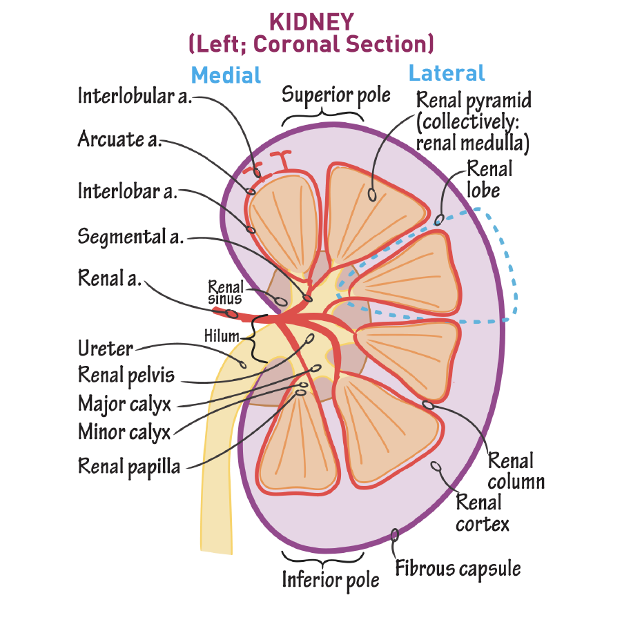
In our second diagram, we show that the renal pelvis forms as a dilation in the ureteric bud. We can now refer to proximal stalk of the ureteric bud as the ureter. In the adult form, the ureter carries urine from the kidney to the bladder, for storage.The ureter exits the kidney at the renal hilum.
At the tips of each new bud, we show mesenchymal caps; cellular interactions between the buds and the caps drive bifurcation.
Ureteric bud branching depends on reciprocal signaling between the epithelium of the ureteric bud and the mesenchymal caps, especially GDNF/RET signaling; WNT 11 is associated with branching tips.
Then, in the metanephric mesoderm, early generations of branching coalesce to form 2-4 major calyces.
In our final diagram, we show that the later generations of ureteric buds also coalesce, forming the minor calyces.
Continued branching ultimately gives rise to collecting ducts, which drain the excretory units. In the adult form, urine travels through the collecting ducts, into the minor, then major, calyces, and through the renal pelvis to the ureter.
For embryological context, we show the lower portion of the embryo at approximately week 5 with the hindgut, cloaca, urogenital sinus, and, where the allantois and vitelline duct (aka, yolk stalk) travel together, the umbilical cord.
We show the intermediate mesoderm and, within the mesoderm, we show the mesonephric ducts and tubules. Notice that, at this stage, the cranial-most tubules have degenerated. Recall that the caudal ends of the mesonephric ducts empty into the urogenital sinus.
Now, show that the ureteric bud arises as an outgrowth of the mesonephric ducts; it grows towards, and ultimately into, the metanephric mesoderm (which is also of intermediate mesoderm origin).
With this early stage established, let’s show how the ureteric bud and metanephric mesoderm interact to form the collecting system, which includes the renal pelvis, calyces, and collecting ducts.
When the ureteric bud meets the metanephric mesoderm, cellular interactions induce a series of bifurcations.
In our second diagram, we show that the renal pelvis forms as a dilation in the ureteric bud. We can now refer to proximal stalk of the ureteric bud as the ureter. In the adult form, the ureter carries urine from the kidney to the bladder, for storage.The ureter exits the kidney at the renal hilum.
At the tips of each new bud, we show mesenchymal caps; cellular interactions between the buds and the caps drive bifurcation.
Ureteric bud branching depends on reciprocal signaling between the epithelium of the ureteric bud and the mesenchymal caps, especially GDNF/RET signaling; WNT 11 is associated with branching tips.
Then, in the metanephric mesoderm, early generations of branching coalesce to form 2-4 major calyces.
In our final diagram, we show that the later generations of ureteric buds also coalesce, forming the minor calyces.
Continued branching ultimately gives rise to collecting ducts, which drain the excretory units. In the adult form, urine travels through the collecting ducts, into the minor, then major, calyces, and through the renal pelvis to the ureter.
 Nnephrogenesis occurs in the metanephric mesoderm around the collecting ducts.
The first stage of nephrogenesis is renal vesicle formation.
We show a single ureteric bud; recognize that this particular bud would be the product of several rounds of bifurcations after the establishment of the calyces.
At the wide end of the ureteric bud, we label the ampulla, and show the mesenchymal cap.
The ureteric bud expresses FGF, LIF, and Wnt9B, which induce mesenchymal cap release of Wnt4.
Cells from the mesenchymal cap condense into a pretubular aggregate, which, driven by Wnt4, will undergo a mesenchymal-to-epithelial transition to form hollow renal vesicles.
In stage 2, to show body formation, we draw a bifurcated ureteric bud. The renal vesicle grows, transitioning from to a “comma-shaped” body to an “S-Shaped” body.
In this same diagram, show that stage 3 is vascularization, which is characterized by endothelial precursors migrating to the proximal ends of the vesicles.
Finally, in stage 4, we show the functional form.
The ureteric bud is now a definitive collecting duct; the nephron drains into it.
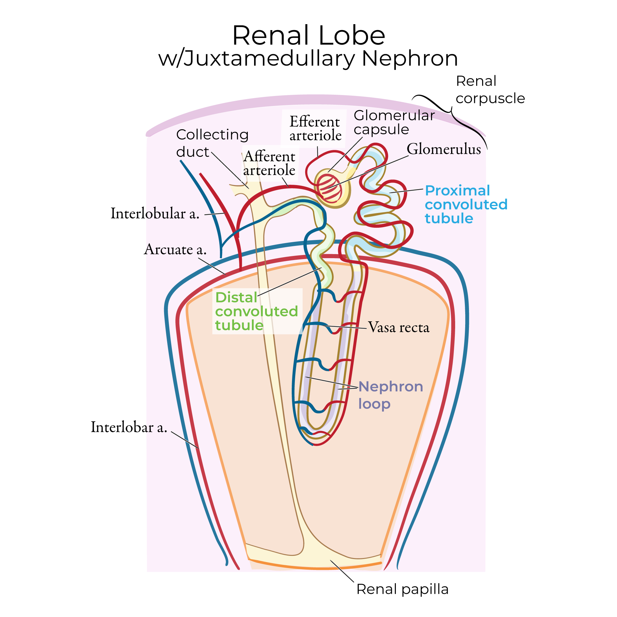
At the proximal end of the nephron, indicate the glomerulus, which is a specialized tuft of capillaries that arose from the endothelial precursors we drew in the vascularization stage.
Surrounding the glomerulus, label the Bowman’s capsule (aka, glomerular capsule).
In the functioning kidney, blood flows through millions of glomeruli, where water, wastes, and other solutes are filtered.
Fetal urine is excreted to the amniotic cavity, which helps maintain the amniotic fluid volume.
Postnatally, the urine content is fine-tuned to maintain acid-base and body water homeostasis as it passes through the nephron; during pregnancy, the placenta performs these functions.
Nnephrogenesis occurs in the metanephric mesoderm around the collecting ducts.
The first stage of nephrogenesis is renal vesicle formation.
We show a single ureteric bud; recognize that this particular bud would be the product of several rounds of bifurcations after the establishment of the calyces.
At the wide end of the ureteric bud, we label the ampulla, and show the mesenchymal cap.
The ureteric bud expresses FGF, LIF, and Wnt9B, which induce mesenchymal cap release of Wnt4.
Cells from the mesenchymal cap condense into a pretubular aggregate, which, driven by Wnt4, will undergo a mesenchymal-to-epithelial transition to form hollow renal vesicles.
In stage 2, to show body formation, we draw a bifurcated ureteric bud. The renal vesicle grows, transitioning from to a “comma-shaped” body to an “S-Shaped” body.
In this same diagram, show that stage 3 is vascularization, which is characterized by endothelial precursors migrating to the proximal ends of the vesicles.
Finally, in stage 4, we show the functional form.
The ureteric bud is now a definitive collecting duct; the nephron drains into it.
At the proximal end of the nephron, indicate the glomerulus, which is a specialized tuft of capillaries that arose from the endothelial precursors we drew in the vascularization stage.
Surrounding the glomerulus, label the Bowman’s capsule (aka, glomerular capsule).
In the functioning kidney, blood flows through millions of glomeruli, where water, wastes, and other solutes are filtered.
Fetal urine is excreted to the amniotic cavity, which helps maintain the amniotic fluid volume.
Postnatally, the urine content is fine-tuned to maintain acid-base and body water homeostasis as it passes through the nephron; during pregnancy, the placenta performs these functions.
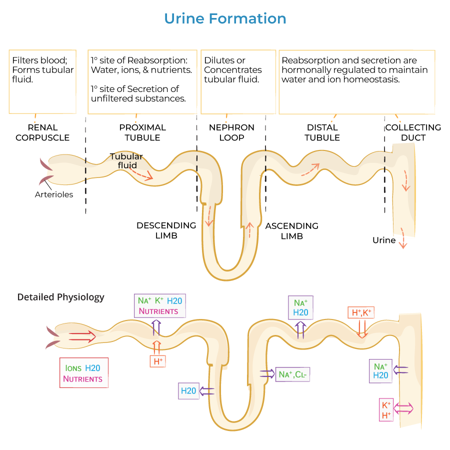 Learn more about Urine Formation
Renal Pelvis, Calyces, and Collecting ducts
Nephrons
Learn more about Urine Formation