

Start your One-Week Free Trial
Already subscribed? Log in »


Development of the Uterus, Uterine Tubes, and Vagina
Here we will learn the embryology of the uterus, uterine tubes, and vagina.
We show the following structures:
The indifferent gonad in approximately week 7, before differentiation of the ovary begins.
The urogenital sinus; recall that this endoderm-derived structure separated from the hindgut earlier in development.
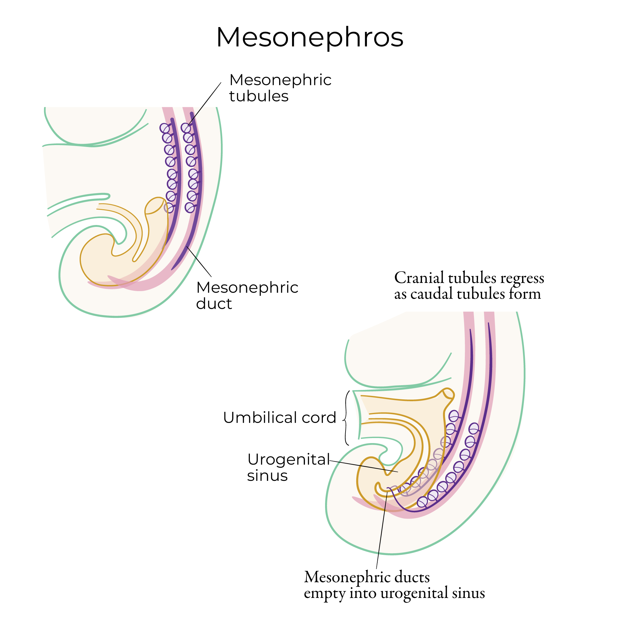
The mesonephric ducts; recall that the mesonephros formed in the intermediate mesoderm as a precursor to the definitive kidney.
 The paramesonephric ducts, which emerged during week 6 as a groove in the coelomic epithelium on the lateral aspect of the urogenital ridge.
The Wnt family of genes is important for the formation and elongation of the paramesonephric ducts in XX embryos (specifically, genes Wnt 4, Wnt 9b, and Wnt 7a).
In approximately weeks 8-10, the paramesonephric ducts partially fuse and give rise to the uterine tubes, uterus, and vagina.
The embryologic origins of the vaginal epithelium is contested:
In the classic dual-origins model, the upper 2/3 of the vaginal epithelium derives from the paramesonephric ducts, and the lower 1/3 derives from the urogenital sinus. In this model, the vaginal epithelium has both mesodermal and endodermal origins (the urogenital sinus has endodermal origins and the paramesonephric ducts has mesodermal origins).
Because this is the model many medical textbooks use, this is the one we'll show in our diagram.
However, be aware that some research, mostly based on mouse studies, indicates a predominantly paramesonephric duct contribution to the vaginal epithelium, but other studies indicate a greater contribution from the urogenital sinus.
The paramesonephric ducts fuse caudally, forming the uterovaginal canal; early in the process, the fusion is incomplete, and a temporary uterine septum is created by the fused paramesonephric ducts.
Caudally, the paramesonephric ducts meet at the sinus tubercle of the urogenital sinus; cranially, the ducts remain open.
The mesonephric ducts regress; at this stage, the ovaries are located between the paramesonephric ducts.
Next, we'll show how the elongation and canalization of the uterus and vagina.
Where the paramesonephric ducts fused caudally, they create the hollow space of the uterine cavity; where the paramesonephric ducts extend and open cranially the uterine tubes (aka fallopian tubes) are taking shape.
Contact between the paramesonephric ducts and the urogenital sinus tubercle induced the formation of the sinovaginal bulbs, which will form the solid vaginal plate.
Continued growth and canalization transform the solid vaginal plate to produce the tube-shaped vagina.
Let's now draw the mature form to see these structures in their final configurations.
Uterus, uterine tubes, and vagina; notice that we've followed the dual-origins model, so the lower vagina has urogenital sinus origins, whereas the rest of the tract developed from the paramesonephric ducts.
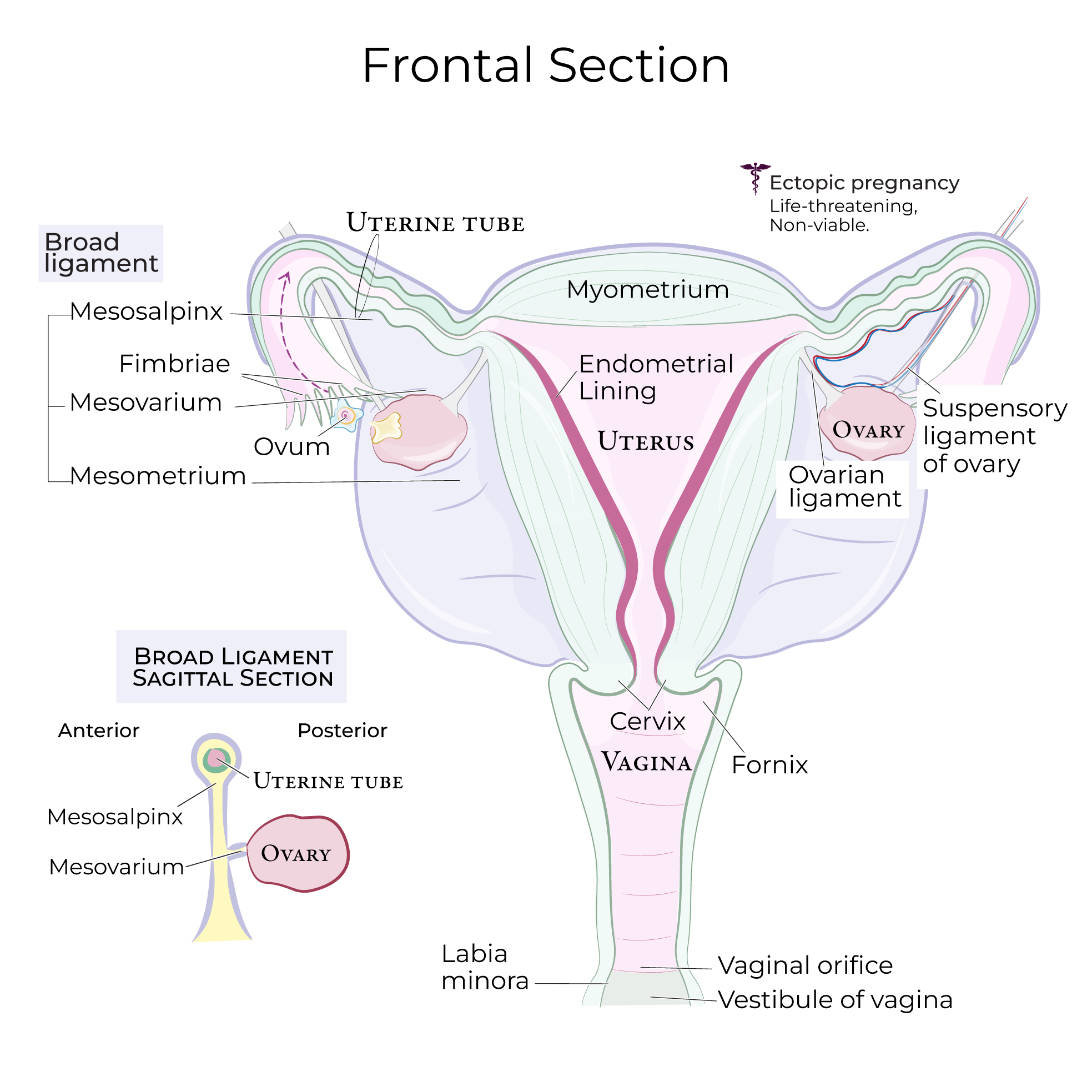
The cervix is the inferior part of the uterus that opens to the vagina, and the hymen, which is a membrane that partially covers the opening of the vagina.
Recall that we left the cranial ends of the paramesonephric ducts open; in the mature form, the fimbriae are the finger-like projections at the open ends of the uterine tubes.
The ovaries descended caudally and laterally from their original locations in the abdomen to their mature locations in the pelvis.
During and after their migration, the ovaries are stabilized by ligaments:
In the mature form, the suspensory ligament of the ovary attaches to the superior pole of the ovary and provides passage for neurovascular structures, and the ovarian ligament anchors the ovary to the uterine wall, adjacent to the attachment of the uterine tubes.
Be aware that other key uterine ligaments include the broad and round ligaments:
The paramesonephric ducts, which emerged during week 6 as a groove in the coelomic epithelium on the lateral aspect of the urogenital ridge.
The Wnt family of genes is important for the formation and elongation of the paramesonephric ducts in XX embryos (specifically, genes Wnt 4, Wnt 9b, and Wnt 7a).
In approximately weeks 8-10, the paramesonephric ducts partially fuse and give rise to the uterine tubes, uterus, and vagina.
The embryologic origins of the vaginal epithelium is contested:
In the classic dual-origins model, the upper 2/3 of the vaginal epithelium derives from the paramesonephric ducts, and the lower 1/3 derives from the urogenital sinus. In this model, the vaginal epithelium has both mesodermal and endodermal origins (the urogenital sinus has endodermal origins and the paramesonephric ducts has mesodermal origins).
Because this is the model many medical textbooks use, this is the one we'll show in our diagram.
However, be aware that some research, mostly based on mouse studies, indicates a predominantly paramesonephric duct contribution to the vaginal epithelium, but other studies indicate a greater contribution from the urogenital sinus.
The paramesonephric ducts fuse caudally, forming the uterovaginal canal; early in the process, the fusion is incomplete, and a temporary uterine septum is created by the fused paramesonephric ducts.
Caudally, the paramesonephric ducts meet at the sinus tubercle of the urogenital sinus; cranially, the ducts remain open.
The mesonephric ducts regress; at this stage, the ovaries are located between the paramesonephric ducts.
Next, we'll show how the elongation and canalization of the uterus and vagina.
Where the paramesonephric ducts fused caudally, they create the hollow space of the uterine cavity; where the paramesonephric ducts extend and open cranially the uterine tubes (aka fallopian tubes) are taking shape.
Contact between the paramesonephric ducts and the urogenital sinus tubercle induced the formation of the sinovaginal bulbs, which will form the solid vaginal plate.
Continued growth and canalization transform the solid vaginal plate to produce the tube-shaped vagina.
Let's now draw the mature form to see these structures in their final configurations.
Uterus, uterine tubes, and vagina; notice that we've followed the dual-origins model, so the lower vagina has urogenital sinus origins, whereas the rest of the tract developed from the paramesonephric ducts.
The cervix is the inferior part of the uterus that opens to the vagina, and the hymen, which is a membrane that partially covers the opening of the vagina.
Recall that we left the cranial ends of the paramesonephric ducts open; in the mature form, the fimbriae are the finger-like projections at the open ends of the uterine tubes.
The ovaries descended caudally and laterally from their original locations in the abdomen to their mature locations in the pelvis.
During and after their migration, the ovaries are stabilized by ligaments:
In the mature form, the suspensory ligament of the ovary attaches to the superior pole of the ovary and provides passage for neurovascular structures, and the ovarian ligament anchors the ovary to the uterine wall, adjacent to the attachment of the uterine tubes.
Be aware that other key uterine ligaments include the broad and round ligaments:
 Finally, let's create a small table to organize the embryological origins of these structures
The paramesonephric ducts give rise to the uterine tubes, uterus, and epithelium of the vagina (or at least to the upper 2/3ds of the epithelium).
The urogenital sinus gives rise to the lower vagina (according to the dual origins model), and, for completion, most of the urinary bladder and urethra.
The cranial genital ligament gives rise to the suspensory ligament of the ovary, and the caudal genital ligament gives rise to the ovarian ligament and the round ligament of the uterus.
Lastly, write that the remnants of the mesonephric tubules include the epoophoron and paroophoron, which are small, non-functional structures sometimes found near the ovary; Gartner's duct, which may become cystic, is a remnant of the mesonephric duct.
Finally, let's create a small table to organize the embryological origins of these structures
The paramesonephric ducts give rise to the uterine tubes, uterus, and epithelium of the vagina (or at least to the upper 2/3ds of the epithelium).
The urogenital sinus gives rise to the lower vagina (according to the dual origins model), and, for completion, most of the urinary bladder and urethra.
The cranial genital ligament gives rise to the suspensory ligament of the ovary, and the caudal genital ligament gives rise to the ovarian ligament and the round ligament of the uterus.
Lastly, write that the remnants of the mesonephric tubules include the epoophoron and paroophoron, which are small, non-functional structures sometimes found near the ovary; Gartner's duct, which may become cystic, is a remnant of the mesonephric duct.
~ Week 7
The paramesonephric ducts, which emerged during week 6 as a groove in the coelomic epithelium on the lateral aspect of the urogenital ridge.
The Wnt family of genes is important for the formation and elongation of the paramesonephric ducts in XX embryos (specifically, genes Wnt 4, Wnt 9b, and Wnt 7a).
~ Weeks 8-10
Mature Form
Summary Table