The lymphatic system is integrated with the immune and cardiovascular systems.
The lymphatic system regulates fluid volume by collecting interstitial fluid and returning it to the systemic circulation as lymphatic fluid. This is achieved by a unidirectional system of vessels that arise in the body's tissues and terminate by emptying into the veins of the neck.
As lymphatic fluid is transported through the lymphatic vessels, specialized organs and tissues monitor the fluid for pathogens. When pathogens are detected, lymphoid cells initiate an immune response.
The lymphatic system also transports molecules that are too large to diffuse through the capillary wall. An example of this is in the small intestine, where specialized lymphatic capillaries, called lacteals, absorb proteins and lipids.
In systemic capillaries, oxygenated blood arrives at the arteriole end and exits at the venule end. Along the length of the capillary, gas exchange occurs via diffusion.
Within the capillary, hydrostatic blood pressure drives filtration, which forces fluid out of the capillary and into the interstitial space; osmotic pressure drives reabsorption of much, but not all, of this fluid.
The remaining fluid, along with proteins and other molecules, are collected by lymphatic capillaries.
Once the interstitial fluid is in the lymphatic system, we call it lymphatic fluid.
Lymphatic fluid is a clear yellowish watery fluid that transports white blood cells, nutrients, and other large molecules within the lymphatic system. As lymph fluid travels through the lymphatic system, it picks up cellular debris and other harmful substances, including pathogens.
B lymphocytes produce antigen-specific antibodies that mark pathogens for destruction.
Cytotoxic T lymphocytes and
natural killer cells directly kill cells that are infected by viruses or are cancerous.
Be aware that there are other T lymphocytes and even more immune cells; you can learn more about these cells and their roles in our notes.
Lymphatic Organs & Tissues
The lymphatic organs and tissues provide the structural basis of the immune system.
The primary lymphoid organs are the bone marrow and the thymus. The thymus is a gland that lies just posterior to your breastbone.
In these primary organs, lymphocytes form and become immunocompetent. Both B and T lymphocytes form in the stem cells of bone marrow; T cells travel to the thymus to complete differentiation.
Lymphocytes leave the primary organs and migrate to secondary organs, where they become activated.
Secondary lymphoid organs include the tonsils, spleen, vermiform appendix, and lymph nodes, which are scattered throughout the body. Mucosa-associated lymphoid tissue (MALT) is also found in various locations; we highlight its presence in the small intestine, where it can also be referred to as GALT (gut-associated lymphoid tissue). Other subtypes of MALT include Bronchus-associated lymphoid tissue and Nasal-associated lymphoid tissue.
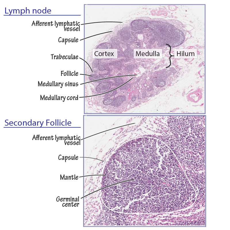
Lymph nodes
Write that lymph nodes house lymphocytes and other immune cells that filter lymph and monitor it for pathogens; they are also the sites of lymphocyte activation. When pathogens are detected, an immune response is initiated to prevent its dissemination.
We show the outer capsule of a lymph node and indicate multiple afferent lymphatic vessels and a single efferent lymphatic vessel.
Lymph fluid flows into the node via the afferent vessels and passes by lymphoid cells organized into outer follicles and inner medullary cords.
As lymph fluid flows past the follicles, the lymphocytes have an opportunity to detect and respond to pathogens.
The lymph fluid exits the node via the efferent lymphatic vessel.
Over 300 lymph nodes drain the head and neck; we highlight the cervical lymph nodes.
Axillary nodes drain the thoracic wall, breast, arm, and the abdominal wall above the umbilicus.
Mediastinal nodes receive lymphatic fluid from the heart, lungs, and other thoracic organs.
Inguinal lymph nodes drain the lower limb and external genitalia.
Popliteal lymph nodes reside in the posterior knee; they drain the leg and foot.
Lmphatic vessels return lymph fluid to the cardiovascular system.
Lymphatic fluid begins its journey in the plexuses of lymphatic capillaries, which drain into larger lymphatic vessels; as we've seen, afferent lymphatic vessels deliver fluid to lymph nodes for filtering.
The efferent lymphatic vessels converge to form lymphatic trunks, which converge again to form two ducts. The two ducts drain into veins of the neck, reintroducing lymphatic fluid to the cardiovascular system.
The right lymphatic duct drains the right sides of the head, neck, and thorax, and the right upper limb (about 25% of the total body lymph fluid).
The thoracic duct has an inferior dilated origin, called the cisterna chyli. The thoracic duct drains the rest of the body (the left sides of the head, neck, thorax, and the left upper limb, as well as the abdomen, pelvis, and both lower limbs).
Unlike the heart in the cardiovascular system, there is no centralized muscular pump to move fluid through the lymphatic system. Instead, the contractions of nearby muscles and the pulsations of nearby arteries push lymph fluid through the lymphatic system; one-way valves prevent backflow, similar to the valves in veins.
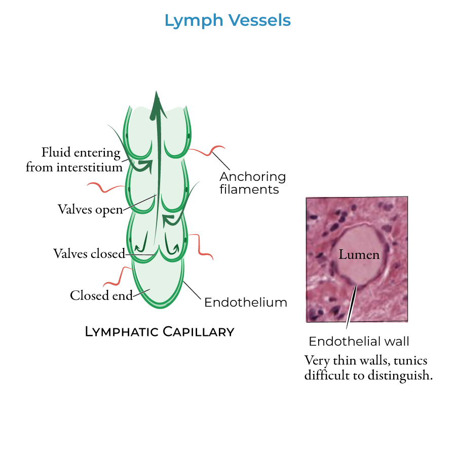
Notice that this is a unidirectional system: lymph fluid is continuously returned to the systemic circulation.
Lymphatic trunks and ducts:
The ducts drain into the right and left brachiocephalic veins, which are formed by the union of the internal jugular and subclavian veins.
The right lymphatic duct returns lymph to the systemic circulation by draining into the right venous angle, which is the junction where the right subclavian and right internal jugular veins merge to form the brachiocephalic vein (be aware that the drainage of the right lymphatic duct is highly variable).
Th thoracic duct begins as distally as the cisterna chyli. The thoracic duct ascends through the thorax and empties into the systemic circulation at the left venous angle.
The trunks are named for the regions of the body they drain.
The paired lumbar lymph trunks drain into the cisterna chyli. The lumbar lymph trunks receive lymph from the lower limbs, pelvis, kidneys and adrenal glands, and the many of the deep lymphatic vessels of the abdominal wall.
An unpaired intestinal trunk also drains into the cisterna chyli; the intestinal trunk drains lymph from the stomach, intestines, pancreas, spleen, and lower part of the liver.
The paired descending thoracic lymph trunks drain into the thoracic duct, which also drains several smaller vessels of the trunk and abdomen.
Superiorly, the thoracic duct drains the left jugular, subclavian, and bronchomediastinal lymph trunks before emptying into the left brachiocephalic vein.
Similarly, on the right side, the right bronchomediastinal, subclavian, and jugular lymph trunks unite at the right lymphatic duct, which drains into the right brachiocephalic vein.
Obstruction in the lymphatic system leads to the accumulation of lymphatic fluid, a condition called lymphedema. Obstruction can occur when the lymphatic vessels or nodes are damaged by surgery, radiation therapy, or injury. In tropical areas, filariasis (threadworm) infection blocks the lymphatic ducts, leading to edema.
A swollen lymph node is called a bubo; buboes are the hallmark of bubonic plague, which is caused by Yersinia pestis bacteria. If not promptly treated with antibiotics, infection spreads to the blood and lungs. You can learn more about this in our notes.


